MyCompetitiveExams Logo MyCompetitiveExams
Search 1,140+ Questions
Chapters, formulas, speed tricks
→
⚡ Brahmastra NEET 2027
720 MARKS

Dynamic 180-question full-length simulator mixed dynamically from all 6 years for 2027 & 2026 aspirants.

Launch Brahmastra 2027 Test →
Start Practice →
100% Free · No Login Required
🌿 Botany 🔥 High-Yield Core Topic (~3–4 Qs/Year)

NEET Structural Organisation in Animals Previous Year Questions with Solutions

Complete repository of official NTA NEET questions for Structural Organisation in Animals from 2020 to 2026. Every question includes 30-second elimination tricks, step-by-step NCERT solutions, option-by-option traps, and official answer keys.

21 Questions 6 Exam Years (2026, 2025, 2024, 2023, 2021, 2020) 17 Easy · 4 Medium · 0 Challenging
NEET 2026 · Q103 Easy
Official NEET 2026 Question 103 - Structural Organisation in Animals
1 skin ✓ Key
2 buccal cavity
3 lungs
4 trachea
💡 Verified Solution & 30-Second Hack
✓
Verified Against NTA Final Answer Key
Option (1) : skin
📚 Structural Organisation in Animals Easy
📋 Given Parameters & Problem Setup
Organism Frog (Rana tigrina, Amphibia)
Environment In water (aquatic respiration)
⚡

The 30-Second Exam Hall Shortcut: ⚡ Frog Respiratory Medium Switch

⏱️ Save ~40 seconds

In water: SKIN only (cutaneous)! On land: Skin, Buccal cavity, and Lungs (pulmonary)!

1 Recall amphibian respiration modes for adult frogs:
2 Cutaneous respiration (via skin): Operates in water, on land, and exclusively during aestivation and hibernation.
3 Buccopharyngeal respiration: Operates on land.
4 Pulmonary respiration (via lungs): Operates on land.
5 In water, the lungs and buccal cavity do NOT function for gas exchange; respiration occurs strictly through the moist, vascularized SKIN.
6 Option (1) is Skin! Done in 5 seconds flat!
🛡️
Why You Can 100% Trust This Shortcut: NCERT Frog Anatomy verbatim: In water, skin acts as aquatic respiratory organ (cutaneous respiration). Dissolved oxygen in the water is exchanged through the skin by diffusion.
📐

Step-by-Step Rigorous Workflow & Mathematical Proof

Step 1

Mechanisms of Respiration in Adult Frogs

Frogs exhibit three distinct modes of respiration depending on their environmental state: 1. Cutaneous Respiration: Gas exchange across the thin, moist, highly vascularized skin. This is the SOLE mode of respiration when the frog is submerged in water, as well as during winter sleep (hibernation) and summer sleep (aestivation). 2. Buccopharyngeal Respiration: Occurs on land when the mouth remains closed while the buccal floor moves up and down. 3. Pulmonary Respiration: Occurs on land using a pair of sac-like lungs.

Respiration in Water≡Cutaneous Respiration (Skin only)\text{Respiration in Water} \equiv \text{Cutaneous Respiration (Skin only)}
  • • Mucus glands keep the skin moist to facilitate diffusion of dissolved oxygen.
Step 2

Evaluate Incorrect Options

1. Buccal cavity: Used only on land; cannot draw dissolved oxygen underwater. 2. Lungs: Frogs cannot breathe water into their lungs; lungs function exclusively in atmospheric air. 3. Trachea: Frogs lack a true trachea (they have a laryngotracheal chamber leading directly to lungs).

Underwater: Lungs and Buccal Cavity are inactive for gas exchange\text{Underwater: Lungs and Buccal Cavity are inactive for gas exchange}
  • • Option (1) is the only scientifically correct medium-specific organ.
Step 3

Conclusion

In water, frogs respire using skin.

Matches Option (1)\text{Matches Option (1)}
  • • Matches Option (1).
⭐

360° Option Elimination Engine Key Feature

Diagnose every choice: Know exactly why the right option works and why distractors fail.

4-Option Deep Scan
1 A
✓ 100% Correct
Verdict & Scientific Reason:

Exact match with NCERT Frog morphology: Cutaneous respiration via skin is the exclusive aquatic respiratory mechanism.

💡
Why this is airtight: None; canonical zoological knowledge.
2 B
✗ Incorrect
Verdict & Scientific Reason:

Incorrect. Buccal respiration occurs only on land in atmospheric air.

⚠️
Student Fallacy (Why candidates pick this trap): Assuming buccal pumping works underwater.
3 C
✗ Incorrect
Verdict & Scientific Reason:

Incorrect. Lungs are used for pulmonary respiration on land, never underwater.

⚠️
Student Fallacy (Why candidates pick this trap): Thinking frogs breathe underwater using lungs.
4 D
✗ Incorrect
Verdict & Scientific Reason:

Incorrect. Frogs do not possess a trachea (tracheae are insect respiratory organs).

⚠️
Student Fallacy (Why candidates pick this trap): Confusing insect respiratory organs with amphibian anatomy.
⚠️

NTA Trap Alert: Confusing Land Respiration with Aquatic Respiration

On land, frogs use Skin + Buccal Cavity + Lungs. In water, frogs use ONLY their skin (Cutaneous respiration).

🛡️ How to avoid the -1 mark: Underwater or sleeping in mud (hibernation/aestivation) = SKIN ONLY.
📖

Core NEET Syllabus Standard

NEET Biology Core Syllabus · Structural Organisation in Animals Standard Benchmark
Core Scientific Principle Tested:

"In water, dissolved oxygen is exchanged across the moist skin by simple diffusion."
Syllabus Focus: Frog anatomy - Respiratory system (Cutaneous, Buccal, Pulmonary)
✓ Core NEET UG Syllabus Anchor Syllabus & Academic Standards ↗
In water or sleeping in mud so deep: Skin is the only lung frogs keep!
NEET 2026 · Q135 Easy
Official NEET 2026 Question 135 - Structural Organisation in Animals
1 6.
2 9
3 10 ✓ Key
4 12
💡 Verified Solution & 30-Second Hack
✓
Verified Against NTA Final Answer Key
Option (3) : 10
📚 Structural Organisation in Animals Easy
📋 Given Parameters & Problem Setup
Organism Indian bullfrog (Rana tigrina)
Anatomical Structure Cranial nerves arising from the brain
Metric Number of PAIRS of cranial nerves
⚡

The 30-Second Exam Hall Shortcut: ⚡ Frog vs Human Cranial Nerve Rule

⏱️ Save ~40 seconds

Frog (Amphibian) = 10 Pairs; Amniotes (Humans/Reptiles/Birds) = 12 Pairs!

1 Recall amphibian cranial anatomy from NCERT Frog chapter:
2 "10 pairs of cranial nerves arise from the brain."
3 Look at Option (3): 10.
4 Beware of Option (4) which is 12 (characteristic of amniotes/humans).
5 For frog, the answer is strictly 10 pairs!
6 Pick Option (3) in 3 seconds.
🛡️
Why You Can 100% Trust This Shortcut: NCERT Biology · Structural Organisation in Animals (Rana tigrina Nervous System: 10 pairs of cranial nerves).
📐

Step-by-Step Rigorous Workflow & Mathematical Proof

Step 1

Anatomy of the Amphibian Nervous System

The nervous system of the frog (Rana tigrina) is organized into a central nervous system (brain and spinal cord), a peripheral nervous system (cranial and spinal nerves), and an autonomic nervous system (sympathetic and parasympathetic).

Peripheral Nervous System=Cranial nerves+Spinal nerves\text{Peripheral Nervous System} = \text{Cranial nerves} + \text{Spinal nerves}
  • • Brain is enclosed in a cartilaginous or bony cranium (brain box).
Step 2

Quantify Cranial Nerves in Frog

In anamniotes (fishes and amphibians including frogs), exactly 10 pairs of cranial nerves arise from the brain (Olfactory I, Optic II, Oculomotor III, Trochlear IV, Trigeminal V, Abducens VI, Facial VII, Auditory VIII, Glossopharyngeal IX, and Vagus X). The 11th (Accessory) and 12th (Hypoglossal) nerves are absent.

Cranial Nerves in Frog=10 pairs\text{Cranial Nerves in Frog} = \mathbf{10\text{ pairs}}
  • • NCERT verbatim: "10 pairs of cranial nerves arise from the brain."
Step 3

Contrast with Amniotes / Humans

In amniotes (reptiles, birds, and mammals including humans), 12 pairs of cranial nerves are present. Candidates who mistakenly apply human anatomy will select 12, which is incorrect for frog.

{Frog (Amphibia):10 pairsHuman (Mammalia):12 pairs\begin{cases} \text{Frog (Amphibia)} &: \mathbf{10\text{ pairs}} \\ \text{Human (Mammalia)} &: 12\text{ pairs} \end{cases}
  • • Option (3) is the correct answer.
⭐

360° Option Elimination Engine Key Feature

Diagnose every choice: Know exactly why the right option works and why distractors fail.

4-Option Deep Scan
1 A
✗ Incorrect
Verdict & Scientific Reason:

Incorrect. Far below the actual count.

⚠️
Student Fallacy (Why candidates pick this trap): Random guess.
2 B
✗ Incorrect
Verdict & Scientific Reason:

Incorrect. Does not represent any vertebrate cranial nerve baseline.

⚠️
Student Fallacy (Why candidates pick this trap): Misremembering nerve numbers.
3 C
✓ 100% Correct
Verdict & Scientific Reason:

100% accurate. Canonical NCERT count of cranial nerve pairs in frog.

💡
Why this is airtight: None; solid amphibian anatomy recall.
4 D
✗ Incorrect
Verdict & Scientific Reason:

Incorrect. 12 pairs are found in amniotes (reptiles, birds, mammals/humans), not in amphibians.

⚠️
Student Fallacy (Why candidates pick this trap): Projecting human cranial anatomy (12 pairs) onto the frog.
⚠️

NTA Trap Alert: Projecting Human Anatomy (12 Pairs) onto Frog (10 Pairs)

Medical aspirants spend extensive time studying the 12 cranial nerves of humans. NTA specifically tests frog cranial nerves to check if students remember that amphibians have only 10 pairs.

🛡️ How to avoid the -1 mark: Frog hops with 10; Humans think with 12!
📖

Core NEET Syllabus Standard

NEET Biology Core Syllabus · Structural Organisation in Animals Standard Benchmark
Core Scientific Principle Tested:

"The peripheral nervous system of the frog comprises 10 pairs of cranial nerves and 10 pairs of spinal nerves."

Syllabus Focus: Anatomy of Frog - Control and Coordination
✓ Core NEET UG Syllabus Anchor Syllabus & Academic Standards ↗
Ten for the Toad in the marshy fen; Twelve for the Human with paper and pen!
NEET 2025 · Q94 Easy
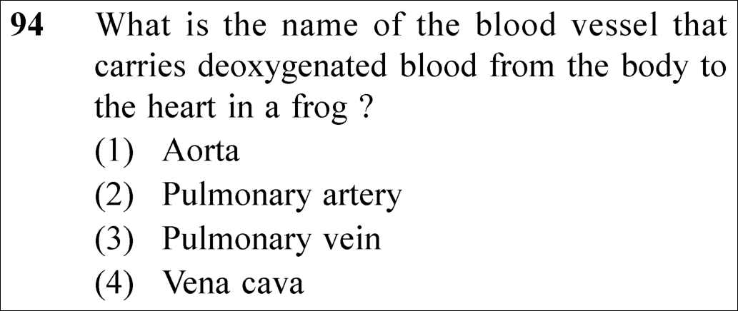
Official NEET 2025 Question 94 - Structural Organisation in Animals
1 Aorta
2 Pulmonary artery
3 Pulmonary vein
4 Vena cava ✓ Key
💡 Verified Solution & 30-Second Hack
✓
Verified Against NTA Final Answer Key
Option (4) : Vena cava
📚 Structural Organisation in Animals Easy
📋 Given Parameters & Problem Setup
Organism Frog (Rana tigrina)
Blood Type Deoxygenated blood
Direction of Flow From body tissues returning to the heart
⚡

The 30-Second Exam Hall Shortcut: ⚡ Amphibian Venous Return Rule

⏱️ Save ~20 seconds

Aorta carries oxygenated blood AWAY from heart. Pulmonary artery carries blood TO lungs. Pulmonary vein carries oxygenated blood FROM lungs. Deoxygenated blood from the body is collected by major veins called VENA CAVA (caval veins) into the sinus venosus! ⟹ Vena cava!

1 Recall frog vascular system: Blood from all body parts is returned to the heart by veins.
2 Major veins from the anterior and posterior body regions are the venae cavae (two precavals and one postcaval).
3 These vena cava vessels pour deoxygenated blood into a triangular sac called sinus venosus, which in turn empties into the right atrium.
4 Contrast with alternatives:
5 - Aorta: Large arterial trunk distributing oxygenated/mixed blood to body tissues.
6 - Pulmonary artery: Carries deoxygenated blood to lungs.
7 - Pulmonary vein: Carries oxygenated blood from lungs to left atrium.
8 Match Option (4) Vena cava in under 5 seconds!
🛡️
Why You Can 100% Trust This Shortcut: Anuran Urogenital Anatomy (NCERT Biology · Structural Organisation in Animals: Ureters act as urinogenital ducts in male frogs).
📐

Step-by-Step Rigorous Workflow & Mathematical Proof

Step 1

Circulatory System of Frog (Rana tigrina)

The vascular system of frog is closed, complete, and contains a three-chambered heart (two atria and one ventricle):

Body Tissues→Deoxygenated bloodVena cava⟶Sinus venosus⟶Right Atrium\text{Body Tissues} \xrightarrow{\text{Deoxygenated blood}} \text{Vena cava} \longrightarrow \text{Sinus venosus} \longrightarrow \text{Right Atrium}
  • • Deoxygenated blood from the head, forelimbs, and body is collected by the anterior and posterior vena cava.
Step 2

Contrast with Other Vessels

Evaluate the physiological role of the other options:

Aorta: Arterial distribution,Pulmonary Vein: Oxygenated return from lungs\text{Aorta: Arterial distribution}, \quad \text{Pulmonary Vein: Oxygenated return from lungs}
  • • Aorta delivers systemic blood outward from the conus arteriosus.
  • • Pulmonary veins carry oxygen-rich blood from the lungs into the left atrium.
  • • Pulmonary arteries transport blood from heart to lungs for oxygenation.
  • • Therefore, the vessel carrying deoxygenated blood from body to heart is the Vena cava, matching Option (4).
⭐

360° Option Elimination Engine Key Feature

Diagnose every choice: Know exactly why the right option works and why distractors fail.

4-Option Deep Scan
1 A
✗ Incorrect
Verdict & Scientific Reason:

Incorrect. The aorta is an artery that transports blood away from the heart to the body.

⚠️
Student Fallacy (Why candidates pick this trap): Confusing arteries with veins.
2 B
✗ Incorrect
Verdict & Scientific Reason:

Incorrect. Carries blood from the heart to the lungs.

⚠️
Student Fallacy (Why candidates pick this trap): Thinking pulmonary artery drains the body.
3 C
✗ Incorrect
Verdict & Scientific Reason:

Incorrect. Carries oxygenated blood from lungs to heart.

⚠️
Student Fallacy (Why candidates pick this trap): Confusing pulmonary vein with systemic veins.
4 D
✓ 100% Correct
Verdict & Scientific Reason:

100% correct. Deoxygenated blood from the body is collected by the vena cava into sinus venosus.

💡
Why this is airtight: None; textbook anatomical recall.
⚠️

NTA Trap Alert: Confusing Sinus Venosus with Vena Cava

Sinus venosus is the receiving heart chamber, whereas VENA CAVA is the actual blood vessel that collects and transports deoxygenated blood from the body into the sinus venosus!

🛡️ How to avoid the -1 mark: The question asks for the "BLOOD VESSEL", which is Vena cava.
📖

Core NEET Syllabus Standard

NEET Biology Core Syllabus · Structural Organisation in Animals Standard Benchmark
Core Scientific Principle Tested:

"Deoxygenated blood from body tissues enters the heart via the vena cava and sinus venosus."

Syllabus Focus: Frog Anatomy: Blood Vascular System
✓ Core NEET UG Syllabus Anchor Syllabus & Academic Standards ↗
Body to Heart = Vena Cava (Vein Caves into heart) ⟹ Option (4)!
NEET 2025 · Q107 Easy
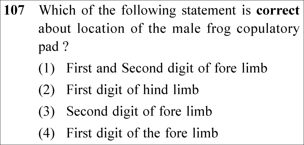
Official NEET 2025 Question 107 - Structural Organisation in Animals
1 First and Second digit of fore limb
2 First digit of hind limb
3 Second digit of fore limb
4 First digit of the fore limb ✓ Key
💡 Verified Solution & 30-Second Hack
✓
Verified Against NTA Final Answer Key
Option (4) : First digit of the fore limb
📚 Structural Organisation in Animals Easy
📋 Given Parameters & Problem Setup
Organism Rana tigrina (Indian Bullfrog)
Structure Copulatory / amplexus pad (nuptial pad)
Sex Male frog during breeding season
⚡

The 30-Second Exam Hall Shortcut: ⚡ Frog Sex Dimorphism Fingerprint

⏱️ Save ~20 seconds

Male frogs have VOCAL SACS (throat) and a COPULATORY PAD on the 1st DIGIT of FORE LIMB! Absent in females!

1 Recall secondary sexual characteristics of male frogs from NCERT:
2 Sound-producing vocal sacs (throat region).
3 Copulatory (nuptial) pad on the FIRST digit of the FORE LIMBS.
4 These pads assist the male in gripping the slippery female during amplexus (mating).
5 Select Option (4) directly!
🛡️
Why You Can 100% Trust This Shortcut: Periplaneta americana Anatomy (NCERT Biology · Structural Organisation in Animals: Hepatic caeca and Malpighian tubules).
📐

Step-by-Step Rigorous Workflow & Mathematical Proof

Step 1

Morphological Sexual Dimorphism in Rana tigrina

Male and female frogs can be distinguished externally during the breeding season by specific dimorphic structures:

  • • Vocal sacs: Present in male frogs below the mouth to amplify mating calls.
  • • Copulatory pad (nuptial pad): A specialised glandular swelling present on the ventral side of the first digit of the forelimb in male frogs.
  • • Females lack both vocal sacs and copulatory pads.
Step 2

Anatomical Position and Function of the Copulatory Pad

During copulatory clasping (amplexus), fertilisation is external in water. The copulatory pad on the 1st digit of the forelimb provides tactile friction and a firm grip on the smooth, mucus-coated body of the female frog.

Step 3

Option Evaluation

The pad is located strictly on the first digit of the forelimb (not on the second digit, and never on the hindlimbs). Thus, Option (4) is 100% correct.

Location=First digit of the fore limb\text{Location} = \text{First digit of the fore limb}
⭐

360° Option Elimination Engine Key Feature

Diagnose every choice: Know exactly why the right option works and why distractors fail.

4-Option Deep Scan
1 A
✗ Incorrect
Verdict & Scientific Reason:

Incorrect. The copulatory pad is restricted exclusively to the first digit of the forelimb, not the second.

⚠️
Student Fallacy (Why candidates pick this trap): Believing the pad spans across multiple digits.
2 B
✗ Incorrect
Verdict & Scientific Reason:

Incorrect. Forelimbs are used for clasping the female; hindlimbs are adapted for swimming and leaping and have no copulatory pad.

⚠️
Student Fallacy (Why candidates pick this trap): Confusing forelimb with hindlimb.
3 C
✗ Incorrect
Verdict & Scientific Reason:

Incorrect. It is present on the first digit, not the second digit.

⚠️
Student Fallacy (Why candidates pick this trap): Misremembering the specific digit number.
4 D
✓ 100% Correct
Verdict & Scientific Reason:

100% correct. NCERT explicitly states copulatory pad is on the first digit of the fore limbs in male frogs.

💡
Why this is airtight: None; direct textbook fact.
⚠️

NTA Trap Alert: Forelimb vs Hindlimb & Digit Number Trap

Examiners frequently swap forelimb with hindlimb, or claim it occurs on the 2nd/3rd digit. Remember: clasping requires the arms (forelimbs) and the "thumb" (1st digit)!

🛡️ How to avoid the -1 mark: Clasping = Front arms (Forelimbs), Thumb = 1st Digit!
📖

Core NEET Syllabus Standard

NEET Biology Core Syllabus · Structural Organisation in Animals Standard Benchmark
Core Scientific Principle Tested:

"Male frogs possess vocal sacs and a copulatory pad on the first digit of the forelimbs."

Syllabus Focus: Frog Morphology · Sexual Dimorphism
✓ Core NEET UG Syllabus Anchor Syllabus & Academic Standards ↗
1st Forearm = 1st digit of Forelimb holds the Female!
NEET 2025 · Q169 Easy
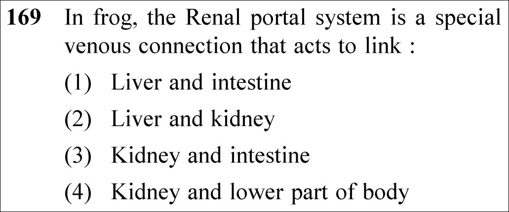
Official NEET 2025 Question 169 - Structural Organisation in Animals
1 Liver and intestine
2 Liver and kidney
3 Kidney and intestine
4 Kidney and lower part of body ✓ Key
💡 Verified Solution & 30-Second Hack
✓
Verified Against NTA Final Answer Key
Option (4) : Kidney and lower part of body
📚 Structural Organisation in Animals Easy
📋 Given Parameters & Problem Setup
Organism Frog (Rana tigrina)
Circulatory Structure Renal portal system
Target Link Organs / body regions linked by this venous portal connection
⚡

The 30-Second Exam Hall Shortcut: ⚡ Frog Portal Systems Fingerprint

⏱️ Save ~15 seconds

Frog has TWO portal systems: Hepatic portal system links Liver and Intestine! Renal portal system links KIDNEY and LOWER PART OF BODY! Option (4)!

1 Recall NCERT Section 7.4.2 (Frog - Vascular System) verbatim:
2 - "Special venous connection between liver and intestine as well as the kidney and lower parts of the body are present in frogs."
3 - "The former is called hepatic portal system and the latter is called renal portal system."
4 - Hepatic portal = Liver and intestine.
5 - Renal portal = Kidney and lower part of body.
6 Target: Renal portal system = Kidney and lower part of body!
7 Select Option (4)!
🛡️
Why You Can 100% Trust This Shortcut: Periplaneta americana Reproductive & Circulatory Systems (NCERT Biology · Structural Organisation in Animals: Spermatophores and mushroom gland).
📐

Step-by-Step Rigorous Workflow & Mathematical Proof

Step 1

Definition of a Portal Venous System

A portal venous system is a vascular arrangement in which blood collected from one capillary bed is conveyed via a portal vein into a second capillary network in an intermediate organ before returning to the heart.

  • • In amphibians such as frogs, two well-developed portal venous circuits exist: the hepatic portal system and the renal portal system.
  • • Humans and mammals retain the hepatic portal system and hypophysial portal system, but the renal portal system is absent (reduced/vestigial).
Step 2

Venous Anatomy of the Frog's Renal Portal System

Tracing the blood vessels in the posterior body:

  • • Deoxygenated venous blood from the hindlimbs and pelvic region is collected by the femoral and sciatic veins.
  • • These bifurcate to form the renal portal veins, which enter the kidneys and break up into a secondary capillary plexus.
  • • This allows the renal tubular epithelium to clear nitrogenous waste and filter blood directly from the lower extremities.
  • • The blood is subsequently collected by renal veins into the postcaval vein (posterior vena cava).
  • • Thus, the renal portal system specifically links the kidney and lower part of the body.
Step 3

Contrast with the Hepatic Portal System

Evaluating Option (1):

  • • The venous connection that links the intestine (alimentary canal) to the liver is the hepatic portal system.
  • • Conflating the hepatic and renal portal systems is an examiner's deliberate trap.
Step 4

Conclusion

The renal portal system acts to link the kidney and lower parts of the body, matching Option (4).

Renal Portal System=Lower part of body→Kidney\text{Renal Portal System} = \text{Lower part of body} \rightarrow \text{Kidney}
⭐

360° Option Elimination Engine Key Feature

Diagnose every choice: Know exactly why the right option works and why distractors fail.

4-Option Deep Scan
1 A
✗ Incorrect
Verdict & Scientific Reason:

Incorrect. The connection between liver and intestine is the hepatic portal system, not the renal portal system.

⚠️
Student Fallacy (Why candidates pick this trap): Confusing hepatic portal system with renal portal system.
2 B
✗ Incorrect
Verdict & Scientific Reason:

Incorrect. There is no direct portal system connecting liver and kidney.

⚠️
Student Fallacy (Why candidates pick this trap): Fabricating a link between two internal visceral organs.
3 C
✗ Incorrect
Verdict & Scientific Reason:

Incorrect. Intestinal veins drain into the hepatic portal system, not the kidney.

⚠️
Student Fallacy (Why candidates pick this trap): Mismatched organ drainage.
4 D
✓ 100% Correct
Verdict & Scientific Reason:

100% correct. Direct verbatim NCERT fact: the renal portal system connects the kidney and lower parts of the body.

💡
Why this is airtight: None; textbook mastery of frog anatomy.
⚠️

NTA Trap Alert: Renal Portal vs Hepatic Portal Systems

Hepatic portal = Intestine -> Liver (carries absorbed food). Renal portal = Hindlimbs / Lower body -> Kidney (carries metabolic waste).

🛡️ How to avoid the -1 mark: Renal = Kidney; connects lower legs/body to the kidney.
📖

Core NEET Syllabus Standard

NEET Biology Core Syllabus · Structural Organisation in Animals Standard Benchmark
Core Scientific Principle Tested:

"In frogs, the renal portal system is a venous connection linking the kidneys with the lower parts of the body."

Syllabus Focus: Frog Anatomy · Vascular System
✓ Core NEET UG Syllabus Anchor Syllabus & Academic Standards ↗
Renal = Running legs to Kidney (Lower body to Kidney)!
NEET 2025 · Q175 Medium
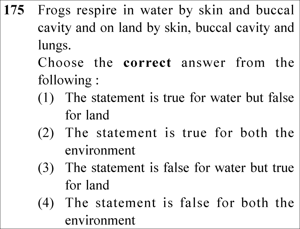
Official NEET 2025 Question 175 - Structural Organisation in Animals
1 The statement is true for water but false for land
2 The statement is true for both the environment
3 The statement is false for water but true for land ✓ Key
4 The statement is false for both the environment
💡 Verified Solution & 30-Second Hack
✓
Verified Against NTA Final Answer Key
Option (3) : The statement is false for water but true for land
📚 Structural Organisation in Animals Medium
📋 Given Parameters & Problem Setup
Proposition Given Frogs respire in water by skin and buccal cavity and on land by skin, buccal cavity and lungs.
Target Evaluation Truth value across both aquatic and terrestrial environments
⚡

The 30-Second Exam Hall Shortcut: ⚡ Frog Respiration Medium Diagnostic

⏱️ Save ~25 seconds

In water, frog respires ONLY by SKIN (cutaneous respiration; mouth and nostrils are tightly sealed)! On land, it respires by SKIN, BUCCAL CAVITY, and LUNGS! Thus, the claim is FALSE for water, but TRUE for land!

1 Check NCERT Section 7.4.2 (Frog - Respiratory System) verbatim:
2 "In water, skin acts as aquatic respiratory organ (cutaneous respiration). Dissolved oxygen in the water is exchanged through the skin by diffusion."
3 - Notice: In water, the buccal cavity does NOT participate (the mouth is closed to prevent water entry into lungs). Thus, claiming "in water by skin and buccal cavity" is FALSE!
4 "On land, the buccal cavity, skin and lungs act as the respiratory organs."
5 - Thus, claiming "on land by skin, buccal cavity and lungs" is TRUE!
6 Conclusion: The statement is false for water but true for land (Option 3)!
7 Select Option (3)!
🛡️
Why You Can 100% Trust This Shortcut: Cockroach Mouthparts & Nervous System Ganglia (NCERT Biology · Structural Organisation in Animals: Biting and chewing type).
📐

Step-by-Step Rigorous Workflow & Mathematical Proof

Step 1

Aquatic Respiration in Rana tigrina (Water Mode)

When submerged underwater or during aestivation/hibernation:

  • • Cutaneous respiration is the exclusive mode of respiration in water.
  • • The skin is highly vascularized, thin, and kept perpetually moist by mucus glands.
  • • Dissolved oxygen in the aquatic medium diffuses directly across the moist epidermis into cutaneous capillaries, while carbon dioxide diffuses out.
  • • The mouth and external nares remain tightly closed underwater; buccal ventilation and pulmonary ventilation do not occur in water.
  • • Therefore, the claim that frogs respire in water by "skin and buccal cavity" is factually FALSE.
Step 2

Terrestrial Respiration in Rana tigrina (Land Mode)

When on land, the frog utilizes three integrated anatomical modalities:

  • • 1. Cutaneous respiration: Oxygen continues to diffuse through the moist skin.
  • • 2. Buccopharyngeal respiration: The lining of the buccal cavity is highly vascular; rhythmic raising and lowering of the hyoid apparatus draws air through external nares into the buccal cavity for gas exchange.
  • • 3. Pulmonary respiration: A pair of elongated, pink, sac-like lungs perform pulmonary ventilation.
  • • Therefore, the claim that on land frogs respire by "skin, buccal cavity and lungs" is completely TRUE.
Step 3

Synthesizing the Final Verdict

Comparing the two clauses of the test statement:

  • • Clause 1 (Water): "Frogs respire in water by skin and buccal cavity" -> FALSE.
  • • Clause 2 (Land): "...and on land by skin, buccal cavity and lungs" -> TRUE.
  • • Hence, the proposition is false for water but true for land.
Step 4

Conclusion

The correct option is Option (3): The statement is false for water but true for land.

Water: Cutaneous ONLY (False)vsLand: Skin + Buccal + Lungs (True)\text{Water: Cutaneous ONLY (False)} \quad \text{vs} \quad \text{Land: Skin + Buccal + Lungs (True)}
⭐

360° Option Elimination Engine Key Feature

Diagnose every choice: Know exactly why the right option works and why distractors fail.

4-Option Deep Scan
1 A
✗ Incorrect
Verdict & Scientific Reason:

Incorrect. Reverses the veracity for both environments.

⚠️
Student Fallacy (Why candidates pick this trap): Thinking buccal cavity works underwater and lungs are absent on land.
2 B
✗ Incorrect
Verdict & Scientific Reason:

Incorrect. Fails to recognize that buccal respiration cannot happen underwater without drowning the respiratory passages.

⚠️
Student Fallacy (Why candidates pick this trap): Assuming the frog uses buccal pumping underwater.
3 C
✓ 100% Correct
Verdict & Scientific Reason:

100% correct. Accurately pinpoints that in water only skin respires, while on land skin, buccal cavity, and lungs all function.

💡
Why this is airtight: None; pristine physiological mastery of amphibian respiration.
4 D
✗ Incorrect
Verdict & Scientific Reason:

Incorrect. The land description is a word-for-word match with NCERT.

⚠️
Student Fallacy (Why candidates pick this trap): Denying the tripartite respiratory mechanism on land.
⚠️

NTA Trap Alert: Water Breathing: Buccal Cavity is INACTIVE

Students often generalize: "frogs have 3 types of respiration, so they use them everywhere". In water, the frog can ONLY use cutaneous respiration (skin). Using the buccal cavity would swallow water into lungs!

🛡️ How to avoid the -1 mark: Water = Skin ONLY. Land = Skin + Buccal + Lungs.
📖

Core NEET Syllabus Standard

NEET Biology Core Syllabus · Structural Organisation in Animals Standard Benchmark
Core Scientific Principle Tested:

"In water, only skin performs respiration; on land, buccal cavity, skin, and lungs act as respiratory organs."

Syllabus Focus: Frog Anatomy · Respiratory System
✓ Core NEET UG Syllabus Anchor Syllabus & Academic Standards ↗
Water = Skin Solo; Land = Trio in Demand (Skin, Buccal, Lungs)!
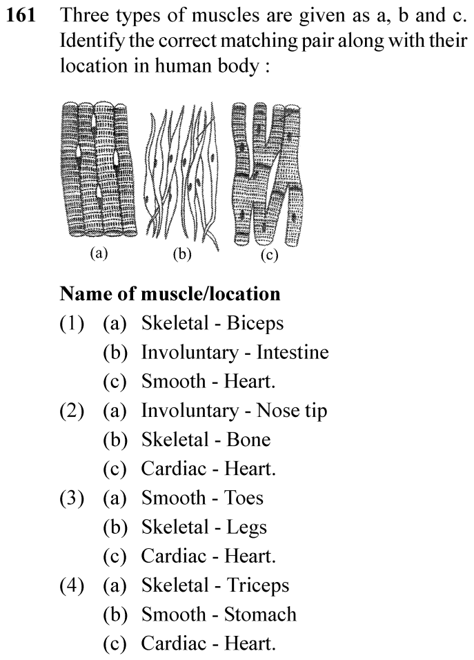
NEET 2024 · Q161 Easy
Official NEET 2024 Question 161 - Structural Organisation in Animals
1 (a) Skeletal - Biceps (b) Involuntary - Intestine (c) Smooth - Heart.
2 (a) Involuntary - Nose tip (b) Skeletal - Bone (c) Cardiac - Heart.
3 (a) Smooth - Toes (b) Skeletal - Legs (c) Cardiac - Heart.
4 (a) Skeletal - Triceps (b) Smooth - Stomach (c) Cardiac - Heart. ✓ Key
💡 Verified Solution & 30-Second Hack
✓
Verified Against NTA Final Answer Key
Option (4) : (a) Skeletal - Triceps; (b) Smooth - Stomach; (c) Cardiac - Heart.
📚 Structural Organisation in Animals Easy
📋 Given Parameters & Problem Setup
Diagram (a) Striated, cylindrical, unbranched fibers with multinucleated peripheral nuclei
Diagram (b) Fusiform, non-striated, single central nucleus
Diagram (c) Striated, branched fibers with intercalated discs
⚡

The 30-Second Exam Hall Shortcut: ⚡ Muscle Histology Visual Signatures

⏱️ Save ~20 seconds

Diagram (a) = Skeletal (striated, unbranched -> Triceps); Diagram (b) = Smooth (spindle-shaped, involuntary -> Stomach); Diagram (c) = Cardiac (branched with intercalated discs -> Heart)!

1 Examine Diagram (a): Parallel cylindrical fibers showing distinct dark/light striations -> Skeletal muscle (e.g., biceps, triceps).
2 Examine Diagram (b): Fusiform / spindle-shaped fibers tapering at both ends, lack striations -> Smooth muscle (visceral organs, e.g., stomach, intestine).
3 Examine Diagram (c): Branched striated fibers with intercalated discs -> Cardiac muscle (heart).
4 Option (4) states: (a) Skeletal - Triceps; (b) Smooth - Stomach; (c) Cardiac - Heart.
5 Select Option (4) / D.
🛡️
Why You Can 100% Trust This Shortcut: NCERT Biology · Structural Organisation in Animals (Muscle Tissue)
📐

Step-by-Step Rigorous Workflow & Mathematical Proof

Step 1

Identify Diagram (a) (Skeletal Muscle)

Skeletal muscle tissue is closely attached to skeletal bones. Striated muscle fibers are bundled together in a parallel fashion with multiple peripheral nuclei. Triceps and biceps are classic examples.

Step 2

Identify Diagram (b) (Smooth Muscle)

Smooth muscle fibers taper at both ends (fusiform) and do not show striations. Cell junctions hold them together and they are bundled in a connective tissue sheath. The wall of internal organs such as blood vessels, stomach and intestine contains this type of muscle tissue.

Step 3

Identify Diagram (c) (Cardiac Muscle)

Cardiac muscle tissue is a contractile tissue present only in the heart. Cell junctions fuse the plasma membranes of cardiac muscle cells and make them stick together. Communication junctions (intercalated discs) at some fusion points allow the cells to contract as a unit.

⭐

360° Option Elimination Engine Key Feature

Diagnose every choice: Know exactly why the right option works and why distractors fail.

4-Option Deep Scan
1 (1)
✗ Incorrect
Verdict & Scientific Reason:

Misidentifies cardiac muscle (c) as smooth muscle.

⚠️
Student Fallacy (Why candidates pick this trap): Misidentifies cardiac muscle (c) as smooth muscle.
2 (2)
✗ Incorrect
Verdict & Scientific Reason:

Skeletal muscles are voluntary; nose tip contains cartilage, not muscle.

⚠️
Student Fallacy (Why candidates pick this trap): Skeletal muscles are voluntary; nose tip contains cartilage, not muscle.
3 (3)
✗ Incorrect
Verdict & Scientific Reason:

Inverts skeletal (a) and smooth (b).

⚠️
Student Fallacy (Why candidates pick this trap): Inverts skeletal (a) and smooth (b).
4 (4)
✓ 100% Correct
Verdict & Scientific Reason:

Correctly identifies the histological morphology and physiological anatomical location for all three muscle types.

💡
Why this is airtight: None.
⚠️

NTA Trap Alert: Intercalated Disc Identification

Confusing cardiac muscle branching with skeletal muscle fibers.

🛡️ How to avoid the -1 mark: Look for dark transverse bands (intercalated discs) and branching patterns unique to cardiac muscle.
📖

Core NEET Syllabus Standard

Biology Core Zoology · Structural Organisation in Animals Standard Benchmark
Core Scientific Principle Tested:

"Skeletal muscle tissue is closely attached to skeletal bones... Smooth muscle fibres taper at both ends and do not show striations. Cardiac muscle tissue is a contractile tissue present only in the heart. Communication junctions (intercalated discs) allow cells to contract as a unit."

Syllabus Focus: Classification of Muscle Tissues: Skeletal, Smooth, and Cardiac
✓ Core NEET UG Syllabus Anchor Syllabus & Academic Standards ↗
NEET 2024 · Q180 Easy
Official NEET 2024 Question 180 - Structural Organisation in Animals
1 8th and 9th segment
2 11 segment
3 5th segment
4 10 segment ✓ Key
💡 Verified Solution & 30-Second Hack
✓
Verified Against NTA Final Answer Key
Option (4) : 10th segment
📚 Structural Organisation in Animals Easy
📋 Given Parameters & Problem Setup
Organism Cockroach (Periplaneta americana)
Appendages Jointed filamentous anal cerci present in BOTH sexes
⚡

The 30-Second Exam Hall Shortcut: ⚡ Cockroach Anal Appendage Fingerprint

⏱️ Save ~15 seconds

Anal CERCI are present in BOTH sexes on the 10th segment! Anal STYLES are present ONLY in males on the 9th sternum!

1 Recall sexual dimorphism and abdominal appendages in cockroach:
2 - In both sexes, the 10th abdominal segment bears a pair of jointed filamentous structures called anal cerci.
3 - In males only, the 9th sternite bears a pair of short, thread-like unjointed anal styles.
4 The question asks for anal cerci in both sexes -> 10th segment.
5 Select Option (4) / D.
🛡️
Why You Can 100% Trust This Shortcut: NCERT Biology · Structural Organisation in Animals (Cockroach Morphology)
📐

Step-by-Step Rigorous Workflow & Mathematical Proof

Step 1

Review Abdominal Segmentation in Cockroach

The abdomen in both male and female cockroach consists of 10 segments.

Step 2

Distinguish Anal Cerci from Anal Styles

NCERT explicitly describes: 1. Anal cerci: In both sexes, the 10th segment bears a pair of jointed filamentous structures called anal cerci (sensory to sound and vibrations). 2. Anal styles: Males bear a pair of short, threadlike anal styles on the 9th sternum which are absent in females.

Step 3

Match with Options

Anal cerci are located on the 10th segment (Option 4).

⭐

360° Option Elimination Engine Key Feature

Diagnose every choice: Know exactly why the right option works and why distractors fail.

4-Option Deep Scan
1 (1)
✗ Incorrect
Verdict & Scientific Reason:

Female genital pouch is formed by 7th, 8th, and 9th sterna.

⚠️
Student Fallacy (Why candidates pick this trap): Female genital pouch is formed by 7th, 8th, and 9th sterna.
2 (2)
✗ Incorrect
Verdict & Scientific Reason:

Cockroach abdomen has only 10 segments in adults.

⚠️
Student Fallacy (Why candidates pick this trap): Cockroach abdomen has only 10 segments in adults.
3 (3)
✗ Incorrect
Verdict & Scientific Reason:

5th segment is an anterior abdominal segment lacking terminal appendages.

⚠️
Student Fallacy (Why candidates pick this trap): 5th segment is an anterior abdominal segment lacking terminal appendages.
4 (4)
✓ 100% Correct
Verdict & Scientific Reason:

Both male and female cockroaches bear jointed filamentous anal cerci on their 10th abdominal segment.

💡
Why this is airtight: None.
⚠️

NTA Trap Alert: Anal Cerci vs Anal Styles Confusion

Confusing 9th segment (anal styles in males only) with 10th segment (anal cerci in both sexes).

🛡️ How to avoid the -1 mark: Cerci = 10th segment (both sexes); Styles = 9th sternum (males only).
📖

Core NEET Syllabus Standard

Biology Core Zoology · Structural Organisation in Animals Standard Benchmark
Core Scientific Principle Tested:

"In both sexes, the 10th segment bears a pair of jointed filamentous structures called anal cerci. Males bear a pair of short, threadlike anal styles which are absent in females."

Syllabus Focus: External Morphology of Cockroach: Anal Cerci Segment Location
✓ Core NEET UG Syllabus Anchor Syllabus & Academic Standards ↗
NEET 2024 · Q192 Easy
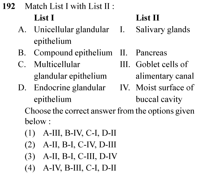
Official NEET 2024 Question 192 - Structural Organisation in Animals
1 ✓ Key
2
3
4
💡 Verified Solution & 30-Second Hack
✓
Verified Against NTA Final Answer Key
Option (1) : A-III, B-IV, C-I, D-II
📚 Structural Organisation in Animals Easy
📋 Given Parameters & Problem Setup
List I (Epithelial Types) A: Unicellular glandular epithelium; B: Compound epithelium; C: Multicellular glandular epithelium; D: Endocrine glandular epithelium
List II (Anatomical Locations / Examples) I: Salivary glands; II: Pancreas; III: Goblet cells of alimentary canal; IV: Moist surface of buccal cavity
⚡

The 30-Second Exam Hall Shortcut: ⚡ Epithelial Tissue Classification Match

⏱️ Save ~25 seconds

Unicellular glands = Goblet cells (III); Compound epithelium = Buccal cavity (protection IV); Multicellular glands = Salivary glands (I); Endocrine glands = Pancreas (islets II)! Match: A-III, B-IV, C-I, D-II!

1 A. Unicellular glandular epithelium: Isolated glandular cells such as Goblet cells of alimentary canal (III).
2 B. Compound epithelium: Multi-layered protective tissue covering moist surface of buccal cavity and pharynx (IV).
3 C. Multicellular glandular epithelium: Cluster of cells forming salivary glands (I).
4 D. Endocrine glandular epithelium: Ductless hormone-secreting tissue, e.g., endocrine pancreas (Islets of Langerhans) (II).
5 Matching order: A-III, B-IV, C-I, D-II -> Option (1) / A.
🛡️
Why You Can 100% Trust This Shortcut: NCERT Biology · Structural Organisation in Animals (Epithelial Tissue Classification)
📐

Step-by-Step Rigorous Workflow & Mathematical Proof

Step 1

Review Glandular Epithelium Classification

Glandular epithelium is classified into two types based on the number of cells: - Unicellular: consisting of isolated glandular cells, like goblet cells of the alimentary canal (A -> III). - Multicellular: consisting of cluster of cells, like salivary glands (C -> I).

Step 2

Review Compound and Endocrine Epithelium

Compound epithelium is made of more than one layer (multi-layered) of cells and thus has a limited role in secretion and absorption. Its main function is to provide protection against chemical and mechanical stresses. They cover the dry surface of the skin, the moist surface of buccal cavity, pharynx, inner lining of ducts of salivary glands and of pancreatic ducts (B -> IV). Endocrine glands do not have ducts; their products called hormones are secreted directly into the fluid bathing the gland, exemplified by the endocrine islets of the pancreas (D -> II).

Step 3

Confirm Match

A-III, B-IV, C-I, D-II corresponds to Option (1).

⭐

360° Option Elimination Engine Key Feature

Diagnose every choice: Know exactly why the right option works and why distractors fail.

4-Option Deep Scan
1 (1)
✓ 100% Correct
Verdict & Scientific Reason:

Accurately pairs all four epithelial configurations with their precise histological locations from NCERT.

💡
Why this is airtight: None.
2 (2)
✗ Incorrect
Verdict & Scientific Reason:

Mismatches Unicellular gland with Pancreas and Compound epithelium with Salivary glands.

⚠️
Student Fallacy (Why candidates pick this trap): Mismatches Unicellular gland with Pancreas and Compound epithelium with Salivary glands.
3 (3)
✗ Incorrect
Verdict & Scientific Reason:

Mismatches Unicellular gland with Pancreas.

⚠️
Student Fallacy (Why candidates pick this trap): Mismatches Unicellular gland with Pancreas.
4 (4)
✗ Incorrect
Verdict & Scientific Reason:

Mismatches Unicellular gland with buccal cavity.

⚠️
Student Fallacy (Why candidates pick this trap): Mismatches Unicellular gland with buccal cavity.
⚠️

NTA Trap Alert: Goblet Cells vs Salivary Glands Cellularity

Confusing unicellular vs multicellular glandular structures.

🛡️ How to avoid the -1 mark: Goblet cells are UNICELLULAR; Salivary glands are MULTICELLULAR.
📖

Core NEET Syllabus Standard

Biology Core Zoology · Structural Organisation in Animals Standard Benchmark
Core Scientific Principle Tested:

"Glandular epithelium is mainly of two types: unicellular, consisting of isolated glandular cells (goblet cells of alimentary canal), and multicellular, consisting of cluster of cells (salivary gland)... Compound epithelium covers the moist surface of buccal cavity."

Syllabus Focus: Classification of Glandular and Compound Epithelial Tissues
✓ Core NEET UG Syllabus Anchor Syllabus & Academic Standards ↗
NEET 2024 · Q200 Easy
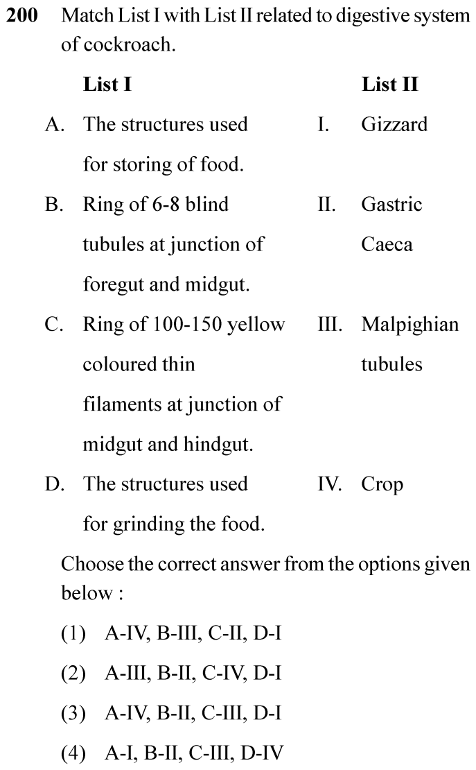
Official NEET 2024 Question 200 - Structural Organisation in Animals
1
2
3 ✓ Key
4
💡 Verified Solution & 30-Second Hack
✓
Verified Against NTA Final Answer Key
Option (3) : A-IV, B-II, C-III, D-I
📚 Structural Organisation in Animals Easy
📋 Given Parameters & Problem Setup
List I (Digestive Structures/Descriptions) A: Structures used for storing food; B: Ring of 6-8 blind tubules at junction of foregut and midgut; C: Ring of 100-150 yellow thin filaments at junction of midgut and hindgut; D: Structures used for grinding food
List II (Anatomical Names) I: Gizzard; II: Gastric Caeca; III: Malpighian tubules; IV: Crop
⚡

The 30-Second Exam Hall Shortcut: ⚡ Cockroach Alimentary Canal Landmark Lock

⏱️ Save ~25 seconds

Food storage = Crop (IV); 6-8 blind tubules at foregut/midgut = Hepatic/Gastric caeca (II); 100-150 yellow tubules at midgut/hindgut = Malpighian tubules (III); Grinding teeth = Gizzard / Proventriculus (I)! Match: A-IV, B-II, C-III, D-I!

1 A. Storing of food: The pharynx opens into a narrow tubular oesophagus which opens into a sac-like structure called Crop (IV).
2 B. Ring of 6-8 blind tubules at junction of foregut and midgut: Hepatic or Gastric Caeca, which secrete digestive juice (II).
3 C. Ring of 100-150 yellow coloured thin filaments at junction of midgut and hindgut: Malpighian tubules, which help in excretion (III).
4 D. Grinding the food: Gizzard (proventriculus) has outer circular muscles and thick inner cuticle forming six chitinous teeth (I).
5 Matching order: A-IV, B-II, C-III, D-I -> Option (3) / C.
🛡️
Why You Can 100% Trust This Shortcut: NCERT Biology · Structural Organisation in Animals (Cockroach Alimentary Canal)
📐

Step-by-Step Rigorous Workflow & Mathematical Proof

Step 1

Review Foregut Organs (Crop and Gizzard)

The oesophagus opens into a sac like structure called crop used for storing of food (A -> IV). The crop is followed by gizzard or proventriculus. It has an outer layer of thick circular muscles and thick inner cuticle forming six highly chitinous plates called teeth. Gizzard helps in grinding the food particles (D -> I).

Step 2

Review Caeca and Excretory Tubules at Gut Junctions

A ring of 6-8 blind tubules called hepatic or gastric caeca is present at the junction of foregut and midgut, which secrete digestive juice (B -> II). At the junction of midgut and hindgut is present another ring of 100-150 yellow coloured thin filamentous Malpighian tubules. They help in removal of excretory products from haemolymph (C -> III).

Step 3

Select Matching Choice

A-IV, B-II, C-III, D-I corresponds to Option (3).

⭐

360° Option Elimination Engine Key Feature

Diagnose every choice: Know exactly why the right option works and why distractors fail.

4-Option Deep Scan
1 (1)
✗ Incorrect
Verdict & Scientific Reason:

Inverts gastric caeca and Malpighian tubules.

⚠️
Student Fallacy (Why candidates pick this trap): Inverts gastric caeca and Malpighian tubules.
2 (2)
✗ Incorrect
Verdict & Scientific Reason:

Mismatches crop with Malpighian tubules.

⚠️
Student Fallacy (Why candidates pick this trap): Mismatches crop with Malpighian tubules.
3 (3)
✓ 100% Correct
Verdict & Scientific Reason:

Accurately pairs all four alimentary canal structures and junctions with their respective physiological names.

💡
Why this is airtight: None.
4 (4)
✗ Incorrect
Verdict & Scientific Reason:

Inverts crop (storing) and gizzard (grinding).

⚠️
Student Fallacy (Why candidates pick this trap): Inverts crop (storing) and gizzard (grinding).
⚠️

NTA Trap Alert: Gastric Caeca vs Malpighian Tubules Position and Count

Confusing the number and junction location of Gastric Caeca (6-8 at foregut/midgut) with Malpighian Tubules (100-150 at midgut/hindgut).

🛡️ How to avoid the -1 mark: Remember: 6-8 blind tubules = Gastric Caeca (secrete enzymes); 100-150 yellow threads = Malpighian Tubules (excretion).
📖

Core NEET Syllabus Standard

Biology Core Zoology · Structural Organisation in Animals Standard Benchmark
Core Scientific Principle Tested:

"Oesophagus opens into a sac like structure called crop used for storing of food. The crop is followed by gizzard... helps in grinding the food particles. A ring of 6-8 blind tubules called hepatic or gastric caeca is present at junction of foregut and midgut... At junction of midgut and hindgut is present another ring of 100-150 yellow coloured Malpighian tubules."

Syllabus Focus: Alimentary Canal of Cockroach: Crop, Gizzard, Hepatic Caeca, and Malpighian Tubules
✓ Core NEET UG Syllabus Anchor Syllabus & Academic Standards ↗
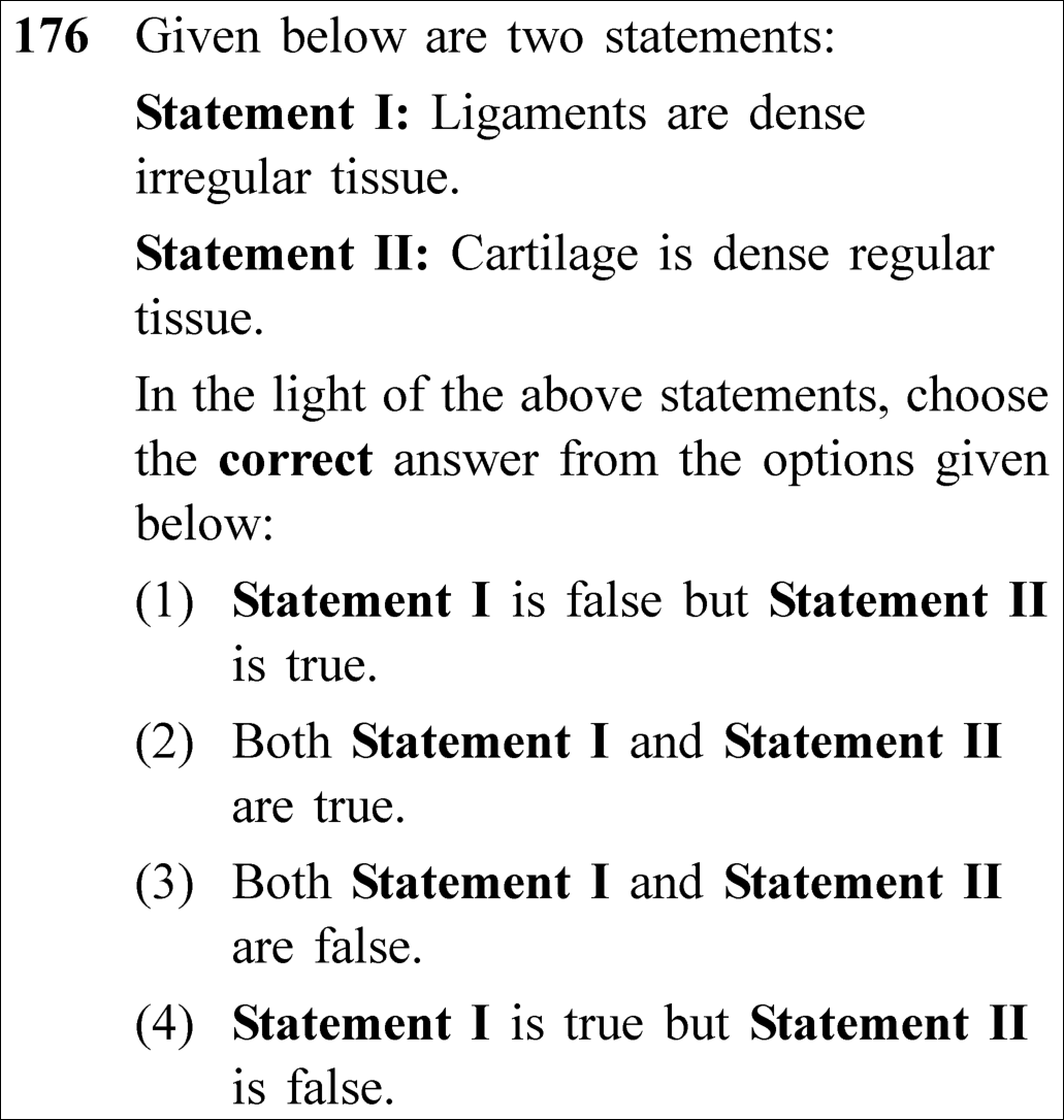
NEET 2023 · Q176 Medium
Official NEET 2023 Question 176 - Structural Organisation in Animals
1 Statement I is false but Statement II is true.
2 Both Statement I and Statement II are true.
3 Both Statement I and Statement II are false. ✓ Key
4 Statement I is true but Statement II is false.
💡 Verified Solution & 30-Second Hack
✓
Verified Against NTA Final Answer Key
Option (3) : Both Statement I and Statement II are false.
📚 Structural Organisation in Animals Medium
📋 Given Parameters & Problem Setup
Statement I Ligaments are dense irregular tissue.
Statement II Cartilage is dense regular tissue.
⚡

The 30-Second Exam Hall Shortcut: ⚡ Connective Tissue Cross-Error: Both are FALSE!

⏱️ Save ~25 seconds

Ligaments and tendons are DENSE REGULAR connective tissue (parallel collagen rows, not irregular!). Cartilage, bone, and blood are SPECIALIZED connective tissues (not dense regular!). Both statements are completely false! Select Option (3)!

1 Dense regular connective tissue: Tendons (muscle to bone) and Ligaments (bone to bone). Statement I calls ligaments dense irregular -> False.
2 Dense irregular connective tissue: Present in skin dermis.
3 Specialized connective tissue: Cartilage, bone, and blood. Statement II calls cartilage dense regular -> False.
4 Both Statement I and Statement II are false.
5 Match Option (3).
🛡️
Why You Can 100% Trust This Shortcut: Canonical NCERT Animal Tissues connective tissue classification matrix.
📐

Step-by-Step Rigorous Workflow & Mathematical Proof

Step 1

Taxonomy of Connective Tissues in NCERT

Connective tissues are divided into three major categories based on ground matrix and fiber density:

Connective Tissue={1. Loose:Areolar, Adipose2. Dense:{Regular:Tendons, LigamentsIrregular:Skin (Dermis)3. Specialised:Cartilage, Bone, Blood\text{Connective Tissue} = \begin{cases} \mathbf{\text{1. Loose:}} & \text{Areolar, Adipose} \\[3pt] \mathbf{\text{2. Dense:}} & \begin{cases} \mathbf{\text{Regular:}} & \mathbf{\text{Tendons, Ligaments}} \\[2pt] \text{Irregular:} & \text{Skin (Dermis)} \end{cases} \\[3pt] \mathbf{\text{3. Specialised:}} & \mathbf{\text{Cartilage, Bone, Blood}} \end{cases}
Step 2

Evaluation of Statement I

Ligaments attach bone to bone and consist of parallel bundles of collagen and elastic fibers arranged in orderly regular rows. Hence, ligaments are dense regular connective tissue. Statement I calls them "dense irregular", which is FALSE.

Ligaments≡Dense Regular Tissue(≠Dense Irregular)  ⟹  Statement I is FALSE\text{Ligaments} \equiv \mathbf{\text{Dense Regular Tissue}} \quad (\neq \text{Dense Irregular}) \implies \text{Statement I is } \mathbf{\text{FALSE}}
Step 3

Evaluation of Statement II

Cartilage has a solid, pliable, chondroitin-rich intercellular material with chondrocytes enclosed in lacunae. NCERT explicitly categorizes cartilage as a specialised connective tissue, alongside bones and blood. Statement II calls it "dense regular", which is FALSE.

Cartilage≡Specialised Connective Tissue(≠Dense Regular)  ⟹  Statement II is FALSE\text{Cartilage} \equiv \mathbf{\text{Specialised Connective Tissue}} \quad (\neq \text{Dense Regular}) \implies \text{Statement II is } \mathbf{\text{FALSE}}
⭐

360° Option Elimination Engine Key Feature

Diagnose every choice: Know exactly why the right option works and why distractors fail.

4-Option Deep Scan
1 1
✗ Incorrect
Verdict & Scientific Reason:

Statement II is false because cartilage is specialized connective tissue, not dense regular.

⚠️
Student Fallacy (Why candidates pick this trap): Thinking cartilage is dense regular connective tissue.
2 2
✗ Incorrect
Verdict & Scientific Reason:

Both statements misclassify their respective connective tissue types.

⚠️
Student Fallacy (Why candidates pick this trap): Assuming both tissue labels are loosely acceptable.
3 3
✓ 100% Correct
Verdict & Scientific Reason:

Both statements are factually false according to canonical NCERT histology classification.

💡
Why this is airtight: None; precise histological taxonomy.
4 4
✗ Incorrect
Verdict & Scientific Reason:

Statement I is false: ligaments are dense regular, not dense irregular.

⚠️
Student Fallacy (Why candidates pick this trap): Believing ligaments possess irregular fiber orientation.
⚠️

NTA Trap Alert: Dense Regular vs. Dense Irregular vs. Specialized Trap

Students frequently remember that tendons and ligaments are dense, but mix up "regular" (parallel bundles) and "irregular" (random criss-cross in skin dermis). Similarly, they forget that cartilage and bone are classified as "specialized".

🛡️ How to avoid the -1 mark: Tendons & Ligaments = Dense Regular. Skin dermis = Dense Irregular. Cartilage, Bone, Blood = Specialized.
📖

Core NEET Syllabus Standard

NCERT Biology · Structural Organisation in Animals Standard Benchmark
Core Scientific Principle Tested:

"Tendons and ligaments are dense regular connective tissue. Cartilage, bones and blood are specialized connective tissues."

Syllabus Focus: Connective Tissue
✓ Core NEET UG Syllabus Anchor Syllabus & Academic Standards ↗
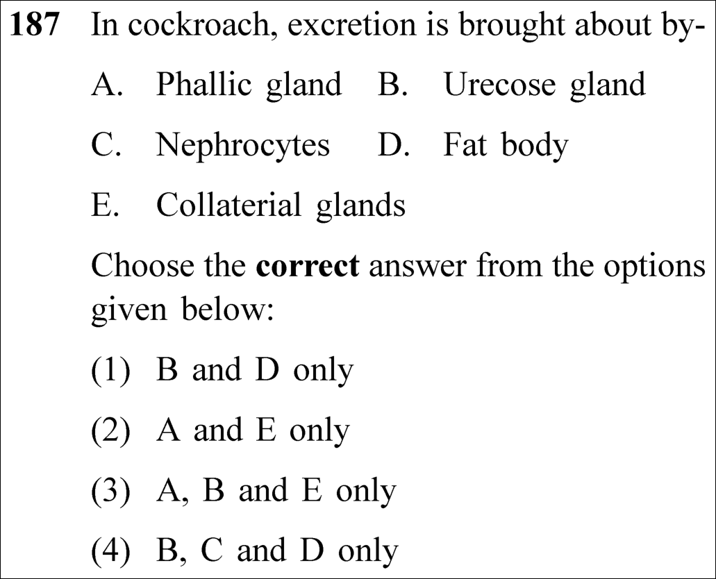
NEET 2023 · Q187 Easy
Official NEET 2023 Question 187 - Structural Organisation in Animals
1 B and D only
2 A and E only
3 A, B and E only
4 B, C and D only ✓ Key
💡 Verified Solution & 30-Second Hack
✓
Verified Against NTA Final Answer Key
Option (4) : B, C and D only
📚 Structural Organisation in Animals Easy
📋 Given Parameters & Problem Setup
Organism Periplaneta americana (Cockroach)
Candidate Structures Phallic gland (A), Urecose gland (B), Nephrocytes (C), Fat body (D), Collaterial glands (E)
⚡

The 30-Second Exam Hall Shortcut: ⚡ Cockroach Excretory Quartet: Tubules, Fat Body, Nephrocytes, Urecose Glands

⏱️ Save ~20 seconds

Phallic gland (A) and Collaterial glands (E) are reproductive structures! Excretion in cockroach is carried out by Malpighian tubules along with Urecose glands (B), Nephrocytes (C), and Fat body (D)! Match Option (4) B, C and D only!

1 Eliminate reproductive structures: Phallic gland (male genital system) and Collaterial glands (female ootheca formation).
2 Retain excretory structures: Urecose gland (B), Nephrocytes (C), Fat body (D).
3 B, C and D only.
4 Match Option (4).
🛡️
Why You Can 100% Trust This Shortcut: Canonical NCERT Cockroach anatomy excretory organ inventory.
📐

Step-by-Step Rigorous Workflow & Mathematical Proof

Step 1

Cockroach Excretory System in NCERT

In cockroach, nitrogenous waste is extracted from haemolymph as potassium urate and converted to uric acid. While the primary excretory organs are Malpighian tubules, accessory excretory structures include:

Excretory Apparatus=Malpighian Tubules+Urecose Glands (B)+Nephrocytes (C)+Fat Body (D)\text{Excretory Apparatus} = \text{Malpighian Tubules} + \mathbf{\text{Urecose Glands (B)}} + \mathbf{\text{Nephrocytes (C)}} + \mathbf{\text{Fat Body (D)}}
Step 2

Verbatim NCERT Reference

NCERT states under Cockroach Excretion: "In addition, the fat body, nephrocytes and urecose glands also help in excretion."

Step 3

Functional Identity of Distractor Structures

Phallic gland (conglobate gland) is an accessory gland of the male reproductive system located near the ejaculatory duct. Collaterial glands are branched glands of the female reproductive system that secrete the scleroprotein hardening the ootheca.

⭐

360° Option Elimination Engine Key Feature

Diagnose every choice: Know exactly why the right option works and why distractors fail.

4-Option Deep Scan
1 1
✗ Incorrect
Verdict & Scientific Reason:

Incomplete: nephrocytes (C) also definitively function in excretion.

⚠️
Student Fallacy (Why candidates pick this trap): Omitting nephrocytes from the auxiliary excretory list.
2 2
✗ Incorrect
Verdict & Scientific Reason:

Both phallic gland (A) and collaterial glands (E) are reproductive structures.

⚠️
Student Fallacy (Why candidates pick this trap): Confusing reproductive accessory glands with excretory organs.
3 3
✗ Incorrect
Verdict & Scientific Reason:

Includes reproductive glands A and E.

⚠️
Student Fallacy (Why candidates pick this trap): Failing to eliminate reproductive organs.
4 4
✓ 100% Correct
Verdict & Scientific Reason:

Verbatim NCERT trio: Urecose gland, nephrocytes, and fat body assist excretion in cockroach.

💡
Why this is airtight: None; textbook verbatim precision.
⚠️

NTA Trap Alert: Reproductive vs. Excretory Glands in Cockroach Trap

Cockroaches have numerous glands ending in "-ic" or "-ial" (phallic, collaterial, urecose, mushroom). Grouping them without noting function leads to errors.

🛡️ How to avoid the -1 mark: Excretory = Urecose gland, Fat body, Nephrocytes. Reproductive = Phallic gland, Collaterial gland.
📖

Core NEET Syllabus Standard

NCERT Biology · Structural Organisation in Animals Standard Benchmark
Core Scientific Principle Tested:

"In addition to Malpighian tubules, the fat body, nephrocytes and urecose glands also help in excretion."

Syllabus Focus: Cockroach (Excretory System)
✓ Core NEET UG Syllabus Anchor Syllabus & Academic Standards ↗
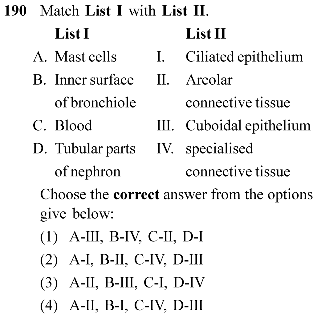
NEET 2023 · Q190 Easy
Official NEET 2023 Question 190 - Structural Organisation in Animals
1
2
3
4 ✓ Key
💡 Verified Solution & 30-Second Hack
✓
Verified Against NTA Final Answer Key
Option (4) : A-II, B-I, C-IV, D-III
📚 Structural Organisation in Animals Easy
📋 Given Parameters & Problem Setup
List I (Cells / Structures) Mast cells, Inner surface of bronchiole, Blood, Tubular parts of nephron
List II (Tissues) Ciliated epithelium, Areolar connective tissue, Cuboidal epithelium, Specialised connective tissue
⚡

The 30-Second Exam Hall Shortcut: ⚡ Histological Location Matcher

⏱️ Save ~25 seconds

Match standard NCERT histological landmarks: Mast cells = Areolar connective tissue (II). Bronchiole lining = Ciliated epithelium (I). Blood = Specialised connective tissue (IV). Nephron tubules = Cuboidal epithelium (III)! Match Option (4)!

1 Mast cells: Resident immune cells in Areolar connective tissue secreting histamine/heparin -> A-II.
2 Inner surface of bronchioles & fallopian tubes: Ciliated epithelium -> B-I.
3 Blood: Fluid specialised connective tissue -> C-IV.
4 Tubular parts of nephrons (PCT, DCT): Cuboidal epithelium -> D-III.
5 Match combination: A-II, B-I, C-IV, D-III -> Option (4).
🛡️
Why You Can 100% Trust This Shortcut: Canonical NCERT Animal Tissues histological classification and location inventory.
📐

Step-by-Step Rigorous Workflow & Mathematical Proof

Step 1

Histological Characteristics of Listed Structures

Analyzing the specific tissue classifications across human body locations:

A. Mast cells:Resident secretory cells in Areolar connective tissue  ⟹  IIB. Bronchiole lining:Ciliated epithelium (propels mucus outward)  ⟹  IC. Blood:Specialised connective tissue (fluid extracellular matrix)  ⟹  IVD. Nephron tubules:Cuboidal epithelium (PCT has microvilli for reabsorption)  ⟹  III\begin{aligned} \text{\textbf{A. Mast cells:}} & \quad \text{Resident secretory cells in } \mathbf{\text{Areolar connective tissue}} \implies \mathbf{\text{II}} \\[4pt] \text{\textbf{B. Bronchiole lining:}} & \quad \mathbf{\text{Ciliated epithelium}} \text{ (propels mucus outward)} \implies \mathbf{\text{I}} \\[4pt] \text{\textbf{C. Blood:}} & \quad \mathbf{\text{Specialised connective tissue}} \text{ (fluid extracellular matrix)} \implies \mathbf{\text{IV}} \\[4pt] \text{\textbf{D. Nephron tubules:}} & \quad \mathbf{\text{Cuboidal epithelium}} \text{ (PCT has microvilli for reabsorption)} \implies \mathbf{\text{III}} \end{aligned}
Step 2

Verbatim NCERT References

NCERT states under Epithelial and Connective Tissues: - "Ciliated epithelium... mainly present in the inner surface of hollow organs like bronchioles and fallopian tubes." - "Cuboidal epithelium is commonly found in ducts of glands and tubular parts of nephrons in kidneys." - "Areolar tissue... contains fibroblasts, macrophages and mast cells." - "Blood is a fluid connective tissue (specialised connective tissue)."

Step 3

Option Cross-Verification

A-II, B-I, C-IV, D-III unambiguously selects Option (4).

⭐

360° Option Elimination Engine Key Feature

Diagnose every choice: Know exactly why the right option works and why distractors fail.

4-Option Deep Scan
1 1
✗ Incorrect
Verdict & Scientific Reason:

Mismatches Mast cells with cuboidal epithelium.

⚠️
Student Fallacy (Why candidates pick this trap): Calling connective tissue cells epithelial cells.
2 2
✗ Incorrect
Verdict & Scientific Reason:

Mismatches Mast cells with ciliated epithelium and bronchioles with areolar tissue.

⚠️
Student Fallacy (Why candidates pick this trap): Swapping mast cells and respiratory linings.
3 3
✗ Incorrect
Verdict & Scientific Reason:

Mismatches bronchioles with cuboidal epithelium and blood with ciliated epithelium.

⚠️
Student Fallacy (Why candidates pick this trap): Scrambling fluid connective tissue with epithelial linings.
4 4
✓ 100% Correct
Verdict & Scientific Reason:

Exact 1-to-1 match with all four canonical NCERT tissue distributions.

💡
Why this is airtight: None; textbook verbatim match.
⚠️

NTA Trap Alert: Bronchioles vs. Nephron Epithelium Trap

Both are tubular hollow organs. Bronchioles have CILIATED epithelium to move mucus; nephron tubules have CUBOIDAL epithelium with microvilli to absorb solutes.

🛡️ How to avoid the -1 mark: Bronchioles = Ciliated. Nephron tubules (PCT/DCT) = Cuboidal.
📖

Core NEET Syllabus Standard

NCERT Biology · Structural Organisation in Animals Standard Benchmark
Core Scientific Principle Tested:

"Mast cells occur in areolar tissue; Bronchioles are lined by ciliated epithelium; Blood is specialized connective tissue; Tubular parts of nephron are lined by cuboidal epithelium."

Syllabus Focus: Animal Tissues
✓ Core NEET UG Syllabus Anchor Syllabus & Academic Standards ↗
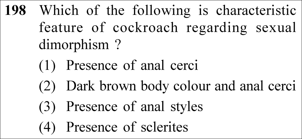
NEET 2023 · Q198 Easy
Official NEET 2023 Question 198 - Structural Organisation in Animals
1 Presence of anal cerci
2 Dark brown body colour and anal cerci
3 Presence of anal styles ✓ Key
4 Presence of sclerites
💡 Verified Solution & 30-Second Hack
✓
Verified Against NTA Final Answer Key
Option (3) : Presence of anal styles
📚 Structural Organisation in Animals Easy
📋 Given Parameters & Problem Setup
Organism Periplaneta americana (Cockroach)
Trait Type Diagnostic feature of sexual dimorphism
⚡

The 30-Second Exam Hall Shortcut: ⚡ Males Have "Style" Rule: Anal Styles in Males Only!

⏱️ Save ~20 seconds

Anal cerci are present in BOTH males and females (on 10th segment, so not dimorphic!). Anal styles (caudal styles) are unjointed thread-like appendages found EXCLUSIVELY in males on the 9th sternite! Match Option (3) Presence of anal styles!

1 Anal cerci: Jointed filamentous sensory structures present on the 10th tergum in BOTH male and female cockroaches.
2 Anal styles: Pair of short, unjointed thread-like structures present on 9th sternite ONLY in males (absent in females).
3 Sclerites and body color are identical in both sexes.
4 Diagnostic sexual dimorphism feature = Presence of anal styles.
5 Match Option (3).
🛡️
Why You Can 100% Trust This Shortcut: Canonical NCERT Cockroach morphology sexual dimorphism criteria.
📐

Step-by-Step Rigorous Workflow & Mathematical Proof

Step 1

Morphological Basis of Sexual Dimorphism in Cockroaches

Male and female cockroaches (Periplaneta americana\textit{Periplaneta americana}) exhibit distinct external abdominal appendages:

Anal Cerci:10th tergite; present in BOTH males and females  ⟹  Not dimorphicAnal Styles:9th sternite; present ONLY in males (absent in females)  ⟹  Diagnostic Sexual Dimorphism\begin{aligned} \text{\textbf{Anal Cerci:}} & \quad \text{10th tergite; present in } \mathbf{\text{BOTH males and females}} \implies \text{Not dimorphic} \\[4pt] \text{\textbf{Anal Styles:}} & \quad \text{9th sternite; present } \mathbf{\text{ONLY in males (absent in females)}} \implies \mathbf{\text{Diagnostic Sexual Dimorphism}} \end{aligned}
Step 2

Verbatim NCERT Reference

NCERT states under Cockroach Morphology: "In both sexes, the 10th segment bears a pair of jointed filamentous structures called anal cerci. In males, genital pouch contains a pair of short, thread-like anal styles which are absent in females."

Step 3

Evaluation of Distractor Traits

Body color (dark brown/reddish brown) and exoskeletal chitinous plates (sclerites: tergites and sternites) are general morphological features shared identically across both sexes.

⭐

360° Option Elimination Engine Key Feature

Diagnose every choice: Know exactly why the right option works and why distractors fail.

4-Option Deep Scan
1 1
✗ Incorrect
Verdict & Scientific Reason:

Anal cerci are present on the 10th segment in both sexes; hence cannot distinguish males from females.

⚠️
Student Fallacy (Why candidates pick this trap): Confusing anal cerci (both sexes) with anal styles (males only).
2 2
✗ Incorrect
Verdict & Scientific Reason:

Neither body color nor anal cerci are sexually dimorphic.

⚠️
Student Fallacy (Why candidates pick this trap): Assuming darker color indicates male gender.
3 3
✓ 100% Correct
Verdict & Scientific Reason:

Verbatim NCERT: unjointed anal styles are present exclusively on the 9th sternum of males and absent in females.

💡
Why this is airtight: None; textbook verbatim recall.
4 4
✗ Incorrect
Verdict & Scientific Reason:

Hardened chitinous sclerites form the exoskeleton of all cockroaches regardless of sex.

⚠️
Student Fallacy (Why candidates pick this trap): Confusing general arthropod anatomy with sexual dimorphism.
⚠️

NTA Trap Alert: Anal Cerci vs. Anal Styles Confusion Trap

Students frequently mix up the two terms: Cerci (found in both, 10th segment) vs. Styles (males only, 9th segment).

🛡️ How to avoid the -1 mark: Use the popular mnemonic: "Males have Style" (Anal styles are unique to males).
📖

Core NEET Syllabus Standard

NCERT Biology · Structural Organisation in Animals Standard Benchmark
Core Scientific Principle Tested:

"In males, the 9th sternite bears a pair of short, thread-like anal styles which are absent in females. Anal cerci are present in both sexes."

Syllabus Focus: Cockroach (Morphology)
✓ Core NEET UG Syllabus Anchor Syllabus & Academic Standards ↗
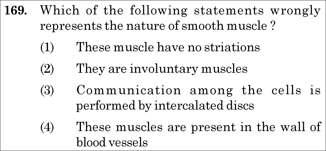
NEET 2021 · Q169 Easy
Official NEET 2021 Question 169 - Structural Organisation in Animals
1 These muscle have no striations
2 They are involuntary muscles
3 Communication among the cells is performed by intercalated discs ✓ Key
4 These muscles are present in the wall of blood vessels
💡 Verified Solution & 30-Second Hack
✓
Verified Against NTA Final Answer Key
Option (3) : Communication among the cells is performed by intercalated discs
📚 Structural Organisation in Animals Easy
📋 Given Parameters & Problem Setup
Tissue Type Smooth Muscle (Non-striated, involuntary)
Question Objective Identify the statement that wrongly represents smooth muscle
⚡

The 30-Second Exam Hall Shortcut: ⚡ "Intercalated Discs are EXCLUSIVE to Cardiac Muscle!"

⏱️ Save ~20 seconds

Intercalated discs (containing gap junctions and desmosomes) are the diagnostic signature of cardiac muscle fibres. Smooth muscles possess simple gap junctions and fusiform cells, never intercalated discs. Option (3) is false for smooth muscle!

1 Check feature: Intercalated discs.
2 Diagnostic truth: Intercalated discs exist ONLY in heart muscle (cardiac muscle).
3 Cross-check other options: Smooth muscle is unstriated (1), involuntary (2), and lines blood vessel walls (4) — all true!
4 Option (3) is the false statement and therefore the correct answer.
🛡️
Why You Can 100% Trust This Shortcut: NCERT Biology · Structural Organisation in Animals (Muscular Tissue: Communication junctions (intercalated discs) at some fusion points allow cardiac cells to contract as a unit; smooth muscles do not have intercalated discs).
📐

Step-by-Step Rigorous Workflow & Mathematical Proof

Step 1

Microscopic and Physiological Profile of Smooth Muscle

Smooth muscle fibres are spindle-shaped (fusiform), unbranched, tapering at both ends, and strictly non-striated (unstriped) because their contractile myofilaments (actin and myosin) are not arranged in regular repeating sarcomeres. - They are involuntary and regulated by the autonomic nervous system. - They form the muscularis coat in the walls of internal visceral organs, including the stomach, intestine, urinary bladder, uterus, and blood vessels (tunica media).

Step 2

Cellular Junctions: Smooth Muscle vs Cardiac Muscle

- In smooth muscle, adjacent fusiform myocytes are bound together by loose connective tissue and communicate via simple gap junctions (nexus) across cell membranes. - In cardiac muscle, adjacent branched fibres are joined end-to-end by specialized zig-zag transverse thickenings called intercalated discs. These structures contain adherens junctions, desmosomes, and gap junctions that synchronize rapid electrical depolarization so the myocardium contracts as a functional syncytium. - Therefore, claiming that smooth muscle cells communicate via intercalated discs is factually wrong.

Intercalated Discs  ⟹  Unique Diagnostic Feature of Cardiac Muscle(NOT Smooth Muscle)\textbf{Intercalated Discs} \implies \text{Unique Diagnostic Feature of } \mathbf{Cardiac\,Muscle} \quad (\text{NOT Smooth Muscle})
⭐

360° Option Elimination Engine Key Feature

Diagnose every choice: Know exactly why the right option works and why distractors fail.

4-Option Deep Scan
1 1
✗ Incorrect
Verdict & Scientific Reason:

This is a true statement for smooth muscle (they lack dark A-bands and light I-bands), so it cannot be the answer to "wrongly represents".

⚠️
Student Fallacy (Why candidates pick this trap): Misreading the negative stem and seeking a true statement.
2 2
✗ Incorrect
Verdict & Scientific Reason:

This is a true statement. Smooth muscles operate automatically without voluntary conscious control.

⚠️
Student Fallacy (Why candidates pick this trap): Confusing voluntary somatic skeletal muscle with involuntary autonomic visceral muscle.
3 3
✓ 100% Correct
Verdict & Scientific Reason:

Correct as the false statement. Intercalated discs are strictly unique to cardiac muscle fibres, never smooth muscle.

💡
Why this is airtight: None. Candidate knows that intercalated discs define cardiac muscle.
4 4
✗ Incorrect
Verdict & Scientific Reason:

This is a true statement. The tunica media of blood vessels is composed of concentric layers of smooth muscle.

⚠️
Student Fallacy (Why candidates pick this trap): Thinking blood vessels contain elastic connective tissue only, unaware of vascular smooth muscle.
⚠️

NTA Trap Alert: Gap Junction vs Intercalated Disc Overlap Trap

Both smooth muscle and cardiac muscle utilize gap junctions for ion transfer. However, intercalated discs are complex mechanical and electrical junctional complexes found *only* at the junctions between cardiac myocytes.

🛡️ How to avoid the -1 mark: Remember: Intercalated disc = Heart / Cardiac muscle only.
📖

Core NEET Syllabus Standard

NEET / NCERT Biology · Structural Organisation in Animals Standard Benchmark
Core Scientific Principle Tested:

"Intercalated discs are specialized junctions found exclusively in cardiac muscle, not smooth muscle."

Syllabus Focus: Muscular Tissue
✓ Core NEET UG Syllabus Anchor Syllabus & Academic Standards ↗
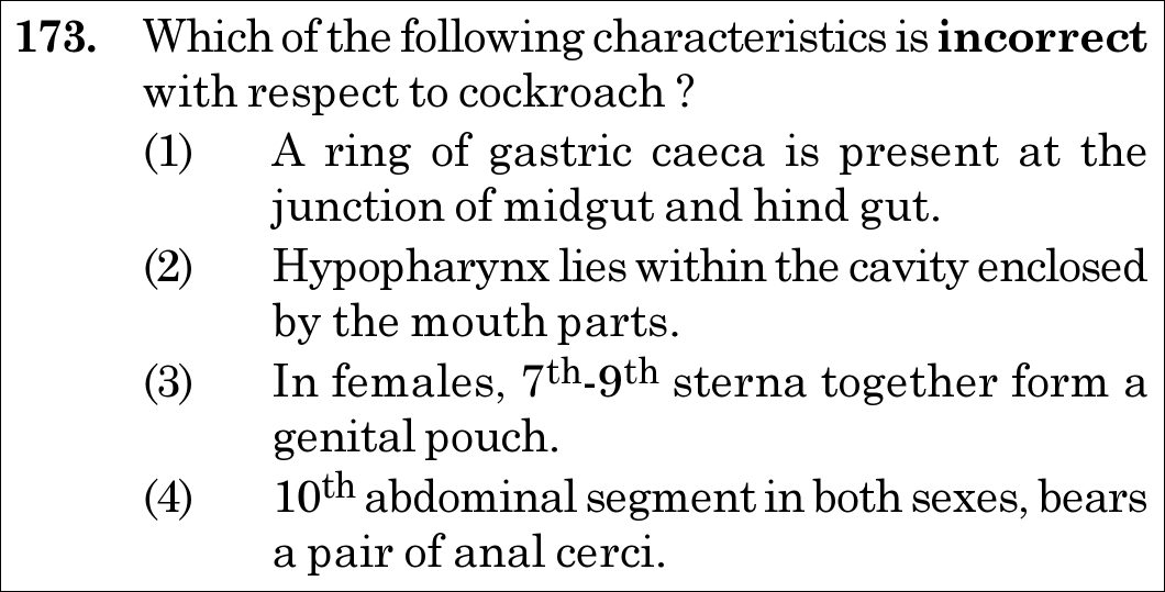
NEET 2021 · Q173 Medium
Official NEET 2021 Question 173 - Structural Organisation in Animals
1 A ring of gastric caeca is present at the junction of midgut and hind gut. ✓ Key
2 Hypopharynx lies within the cavity enclosed by the mouth parts.
3 In females, 7th-9th sterna together form a genital pouch.
4 10th abdominal segment in both sexes, bears a pair of anal cerci.
💡 Verified Solution & 30-Second Hack
✓
Verified Against NTA Final Answer Key
Option (1) : A ring of gastric caeca is present at the junction of midgut and hind gut.
📚 Structural Organisation in Animals Medium
📋 Given Parameters & Problem Setup
Organism Cockroach (Periplaneta americana)
Question Objective Identify the INCORRECT anatomical characteristic
⚡

The 30-Second Exam Hall Shortcut: ⚡ "Gastric Caeca at Foregut-Midgut, Malpighian Tubules at Midgut-Hindgut"

⏱️ Save ~25 seconds

Gastric (hepatic) caeca (6–8 blind tubules) are located at the junction of FOREGUT and MIDGUT (secreting digestive juice). At the junction of MIDGUT and HINDGUT, there are 100–150 yellow Malpighian tubules! Option (1) inverts this boundary and is INCORRECT!

1 Check gut boundaries: - Junction of Foregut (gizzard) & Midgut: 6–8 blind hepatic/gastric caeca. - Junction of Midgut & Hindgut (ileum): 100–150 thin yellow Malpighian tubules.
2 Evaluate Option (1): Claims gastric caeca are at midgut-hindgut junction -> Factually FALSE!
3 Verify others: Hypopharynx in mouth cavity (true), 7th-9th sterna form genital pouch in females (true), anal cerci on 10th segment in both sexes (true).
4 Option (1) is the incorrect characteristic and therefore the correct answer.
🛡️
Why You Can 100% Trust This Shortcut: NCERT Biology · Structural Organisation in Animals (Cockroach Alimentary Canal: A ring of 6-8 blind tubules called hepatic or gastric caeca is present at the junction of foregut and midgut; Malpighian tubules are at the junction of midgut and hindgut).
📐

Step-by-Step Rigorous Workflow & Mathematical Proof

Step 1

Anatomy of the Cockroach Alimentary Canal

The digestive tract of *Periplaneta americana* is divided into three distinct morphological regions: - Foregut (Stomodaeum): Comprises mouth, pharynx, oesophagus, crop (food storage), and gizzard / proventriculus (cuticular teeth for grinding). - Junction of Foregut and Midgut: Possesses a ring of 6–8 blind tubules termed hepatic or gastric caeca, which secrete digestive juices into the mesenteron. - Midgut (Mesenteron): True secretory and absorptive epithelial stomach. - Junction of Midgut and Hindgut: Possesses a ring of 100–150 yellow-coloured, thin filamentous Malpighian tubules, which perform excretion by extracting uric acid and potassium urate from the haemolymph. - Hindgut (Proctodaeum): Differentiated into ileum, colon, and rectum.

{Foregut-Midgut Junction:6–8 Gastric (Hepatic) CaecaMidgut-Hindgut Junction:100–150 Malpighian Tubules\begin{cases} \textbf{Foregut-Midgut Junction:} & 6\text{--}8 \text{ Gastric (Hepatic) Caeca} \\ \textbf{Midgut-Hindgut Junction:} & 100\text{--}150 \text{ Malpighian Tubules} \end{cases}
Step 2

Verification of Remaining Morphological Statements

- Option (2): The hypopharynx is a median flexible lobe acting as the tongue, situated within the preoral cavity enclosed by labrum, mandibles, maxillae, and labium (True). - Option (3): In females, the 7th sternum is boat-shaped and, together with the 8th and 9th sterna, forms a female genital pouch (gynatrium) enclosing the gonopore, spermathecal pores, and collateral glands (True). - Option (4): The 10th abdominal segment in both males and females bears a pair of jointed filamentous sensory structures called anal cerci (True; while anal styles are present only on the 9th sternum of males).

⭐

360° Option Elimination Engine Key Feature

Diagnose every choice: Know exactly why the right option works and why distractors fail.

4-Option Deep Scan
1 1
✓ 100% Correct
Verdict & Scientific Reason:

Correct as the incorrect statement. Gastric caeca are present at the junction of foregut and midgut, not midgut and hindgut.

💡
Why this is airtight: None. Candidate notices the classic NTA anatomical boundary inversion.
2 2
✗ Incorrect
Verdict & Scientific Reason:

This is a true statement describing the cockroach tongue / hypopharynx.

⚠️
Student Fallacy (Why candidates pick this trap): Thinking the hypopharynx is an external sensory antenna.
3 3
✗ Incorrect
Verdict & Scientific Reason:

This is a true statement. The 7th, 8th, and 9th sterna form the female brood pouch / genital chamber.

⚠️
Student Fallacy (Why candidates pick this trap): Confusing male genital pouch (9th and 10th terga with 9th sternum) with female pouch (7th-9th sterna).
4 4
✗ Incorrect
Verdict & Scientific Reason:

This is a true statement. Anal cerci are present on the 10th segment in both males and females.

⚠️
Student Fallacy (Why candidates pick this trap): Confusing anal cerci (both sexes, 10th segment) with anal styles (males only, 9th sternum).
⚠️

NTA Trap Alert: Caeca vs Tubules Boundary Inversion Trap

NTA routinely swaps the junction sites: Gastric caeca (foregut-midgut) vs Malpighian tubules (midgut-hindgut). Hasty readers see "gastric caeca... junction" and assume it is correct without checking which two gut regions are named.

🛡️ How to avoid the -1 mark: Always recite: "Caeca are upstream (foregut-midgut); Malpighian tubules are downstream (midgut-hindgut)".
📖

Core NEET Syllabus Standard

NEET / NCERT Biology · Structural Organisation in Animals Standard Benchmark
Core Scientific Principle Tested:

"Hepatic or gastric caeca are located at the junction of foregut and midgut, while Malpighian tubules are at the junction of midgut and hindgut."

Syllabus Focus: Cockroach Digestive Anatomy
✓ Core NEET UG Syllabus Anchor Syllabus & Academic Standards ↗
NEET 2021 · Q194 Easy
Official NEET 2021 Question 194 - Structural Organisation in Animals
1 Gap junctions and Adhering junctions, respectively.
2 Tight junctions and Gap junctions, respectively. ✓ Key
3 Adhering junctions and Tight junctions, respectively.
4 Adhering junctions and Gap junctions, respectively.
💡 Verified Solution & 30-Second Hack
✓
Verified Against NTA Final Answer Key
Option (2) : Tight junctions and Gap junctions, respectively.
📚 Structural Organisation in Animals Easy
📋 Given Parameters & Problem Setup
Function 1 Stop leakage of substances across a tissue
Function 2 Facilitate communication with neighbouring cells via rapid transfer of ions and small molecules
Question Objective Identify the respective cell junction types
⚡

The 30-Second Exam Hall Shortcut: ⚡ "Stop Leakage = TIGHT Junctions; Rapid Transfer = GAP Junctions"

⏱️ Save ~20 seconds

Tight junctions create a leak-proof barrier stopping paracellular leakage across epithelial sheets. Gap junctions form hydrophilic transmembrane channels (connexons) facilitating rapid transfer of ions and small molecules. In chronological order: Tight junctions and Gap junctions! Select Option (2).

1 Recall NCERT 3 epithelial cell junctions: - Tight junctions: Help stop substances from leaking across a tissue. - Adhering junctions: Perform cementing to keep neighbouring cells together. - Gap junctions: Facilitate the cells to communicate with each other by connecting the cytoplasm for rapid transfer of ions, small molecules, and sometimes big molecules.
2 Match Function 1 ("stop leakage"): Tight junctions.
3 Match Function 2 ("facilitation of communication"): Gap junctions.
4 Sequence required: Tight junctions and Gap junctions, respectively -> Option (2).
🛡️
Why You Can 100% Trust This Shortcut: NCERT Biology · Structural Organisation in Animals (Epithelial Tissue: Tight junctions help to stop substances from leaking across a tissue. Adhering junctions perform cementing to keep neighbouring cells together. Gap junctions facilitate the cells to communicate with each other by connecting the cytoplasm of adjoining cells, for rapid transfer of ions, small molecules and sometimes big molecules).
📐

Step-by-Step Rigorous Workflow & Mathematical Proof

Step 1

Morphological and Molecular Profiles of Epithelial Junctions

Epithelial tissues line body surfaces, cavities, and tubules, providing protective and barrier functions. Adjacent epithelial cells are interlinked by three specialized junctional complexes: - 1. Tight Junctions (Zonula Occludens): Transmembrane claudin and occludin proteins fuse adjacent plasma membranes into a continuous impermeable belt, preventing the paracellular leakage of luminal fluids, ions, and solutes across the tissue. - 2. Adhering Junctions (Zonula Adherens / Desmosomes): Cadherin adhesion molecules anchored to actin microfilaments or intermediate keratin filaments cement adjacent cells together, providing mechanical tensile strength. - 3. Gap Junctions (Nexus): Hexameric rings of connexin proteins form hollow cylindrical channels (connexons) spanning the intercellular gap, directly coupling the cytoplasms of adjacent cells to permit the rapid, unhindered transfer of inorganic ions, second messengers (cAMP, IP₃), metabolites, and small molecules.

Stop Leakage  ⟺  Tight JunctionsRapid Communication  ⟺  Gap JunctionsMechanical Cementing  ⟺  Adhering Junctions\begin{aligned} \textbf{Stop Leakage} &\iff \mathbf{Tight\,Junctions} \\[4pt] \textbf{Rapid Communication} &\iff \mathbf{Gap\,Junctions} \\[4pt] \textbf{Mechanical Cementing} &\iff \mathbf{Adhering\,Junctions} \end{aligned}
Step 2

Positional and Sequential Alignment

The question specifically requires the respective pairing for (1) stopping leakage and (2) communicating via ion transfer. This corresponds strictly to "Tight junctions and Gap junctions, respectively" (Option 2).

⭐

360° Option Elimination Engine Key Feature

Diagnose every choice: Know exactly why the right option works and why distractors fail.

4-Option Deep Scan
1 1
✗ Incorrect
Verdict & Scientific Reason:

Incorrect. Assigns gap junctions to stopping leakage and adhering junctions to communication, which is entirely backwards.

⚠️
Student Fallacy (Why candidates pick this trap): Reversing the physiological functions of junctions.
2 2
✓ 100% Correct
Verdict & Scientific Reason:

Correct. Accurately pairs stopping leakage with tight junctions and ionic communication with gap junctions in the exact requested order.

💡
Why this is airtight: None. Candidate recognizes verbatim NCERT functional definitions.
3 3
✗ Incorrect
Verdict & Scientific Reason:

Incorrect. Adhering junctions perform mechanical cementing, not leakage prevention, and tight junctions do not facilitate intercellular communication.

⚠️
Student Fallacy (Why candidates pick this trap): Confusing mechanical adhering with sealing barriers.
4 4
✗ Incorrect
Verdict & Scientific Reason:

Incorrect because the first function (stopping leakage) is performed by tight junctions, not adhering junctions.

⚠️
Student Fallacy (Why candidates pick this trap): Assuming adhering junctions prevent fluid leakage.
⚠️

NTA Trap Alert: Order and Function Transposition Trap

Candidates sometimes remember the three types of junctions but overlook the order ("respectively") or confuse "cementing" (adhering) with "sealing/stopping leakage" (tight).

🛡️ How to avoid the -1 mark: Use keyword association: "Tight = Leak-proof Seal", "Adhering = Glue/Cement", "Gap = Communicating Bridge".
📖

Core NEET Syllabus Standard

NEET / NCERT Biology · Structural Organisation in Animals Standard Benchmark
Core Scientific Principle Tested:

"Tight junctions prevent substance leakage; gap junctions facilitate rapid ion/molecule transfer between neighbouring cells."

Syllabus Focus: Epithelial Tissue Cell Junctions
✓ Core NEET UG Syllabus Anchor Syllabus & Academic Standards ↗
NEET 2021 · Q200 Medium
Official NEET 2021 Question 200 - Structural Organisation in Animals
1 (a), (b) and (c) are correct ✓ Key
2 (a), (b) and (d) are correct
3 (a), (b), (c) and (d) are correct
4 (b) and (c) are correct - o 0 o -
💡 Verified Solution & 30-Second Hack
✓
Verified Against NTA Final Answer Key
Option (1) : (a), (b) and (c) are correct
📚 Structural Organisation in Animals Medium
📋 Given Parameters & Problem Setup
Anatomical Structure Prostomium of Earthworm (Pheretima posthuma)
Statement (a) It serves as a covering for mouth
Statement (b) It helps to open cracks in the soil into which it can crawl
Statement (c) It is one of the sensory structures
Statement (d) It is the first body segment
⚡

The 30-Second Exam Hall Shortcut: ⚡ "PERISTOMIUM is the First Segment; PROSTOMIUM is the Sensory Upper Lip Wedge!"

⏱️ Save ~25 seconds

The first body segment of the earthworm is the PERISTOMIUM (buccal segment). The prostomium is NOT a body segment; it is a pre-segmental fleshy lobe acting as a mouth covering, a soil-wedge, and a sensory organ. Statement (d) is FALSE! Discard any option with (d) -> Options 2 and 3 vanish. Option (1) is the correct answer!

1 Critical Anatomical Distinction: - Prostomium = Pre-segmental sensory lobe overhang serving as mouth covering and wedge. - Peristomium = True 1st body segment containing the ventral mouth.
2 Evaluate Statement (d): "It is the first body segment" -> Factually FALSE (peristomium is the 1st segment).
3 Eliminate options containing (d): Disqualifies Option (2) and Option (3).
4 Evaluate remaining statements: (a) mouth covering (True), (b) wedges cracks in soil (True), (c) sensory in function (True).
5 Option (1) is 100% correct!
🛡️
Why You Can 100% Trust This Shortcut: NCERT Biology · Structural Organisation in Animals (Earthworm Morphology: The anterior end consists of the mouth and the prostomium, a lobe which serves as a covering for the mouth and as a wedge to open cracks in the soil into which the earthworm may crawl. The prostomium is sensory in function. The first body segment is called the peristomium (buccal segment) which contains the mouth).
📐

Step-by-Step Rigorous Workflow & Mathematical Proof

Step 1

Anatomy of the Anterior Region of Pheretima posthuma

The anterior cephalic end of the earthworm features specialized adaptations for subterranean burrowing in soil: - Prostomium: A fleshy, muscular dorsal lobe projecting above and overhanging the ventral mouth opening. It is pre-segmental (not a true metamere / body segment): - (a) Mouth Covering: It acts as a protective dorsal hood over the mouth. - (b) Soil Wedging: It functions as a powerful mechanical wedge to force open crevices and fissures in compact soil, allowing the worm to crawl forward. - (c) Sensory Function: It is heavily innervated with photoreceptors and chemoreceptive tactile cells to sample light, chemical gradients, and soil moisture.

Step 2

Dissection of Statement (d): Prostomium vs Peristomium

- Peristomium (Buccal Segment): The first true body segment of the earthworm is called the peristomium, which completely encircles the crescentic mouth opening. - Because the prostomium is merely a pre-segmental fleshy lobe overhanging segment 1, stating that the prostomium is the first body segment ((d)) is a direct anatomical error. - Therefore, statements (a), (b), and (c) are correct, while statement (d) is incorrect.

{Prostomium:Pre-segmental Sensory Wedge & Mouth Covering [(a), (b), (c) True]Peristomium:First True Body Segment (Buccal Segment)  ⟹  [(d) is False]\begin{cases} \textbf{Prostomium:} & \text{Pre-segmental Sensory Wedge \& Mouth Covering } [\text{(a), (b), (c) True}] \\[4pt] \textbf{Peristomium:} & \mathbf{First\,True\,Body\,Segment\,(Buccal\,Segment)} \implies [\text{(d) is False}] \end{cases}
⭐

360° Option Elimination Engine Key Feature

Diagnose every choice: Know exactly why the right option works and why distractors fail.

4-Option Deep Scan
1 1
✓ 100% Correct
Verdict & Scientific Reason:

Correct. Confirms all three genuine functions of the prostomium while correctly excluding the false statement (d).

💡
Why this is airtight: None. Candidate knows the distinction between prostomium and peristomium.
2 2
✗ Incorrect
Verdict & Scientific Reason:

Incorrect because (d) is false; peristomium is the first body segment.

⚠️
Student Fallacy (Why candidates pick this trap): Confusing prostomium with peristomium.
3 3
✗ Incorrect
Verdict & Scientific Reason:

Incorrect due to the false inclusion of (d).

⚠️
Student Fallacy (Why candidates pick this trap): Assuming all four statements are true without checking the definition of peristomium.
4 4
✗ Incorrect
Verdict & Scientific Reason:

Incorrect because statement (a) is also a genuine verified function (serves as a covering for the mouth).

⚠️
Student Fallacy (Why candidates pick this trap): Failing to realize that prostomium covers the mouth.
⚠️

NTA Trap Alert: Prostomium vs Peristomium Terminology Trap

NTA intentionally uses "Prostomium" and includes "first body segment" as statement (d). Candidates who confuse the prefixes "Pro-" (before) and "Peri-" (around) fail to realize that *peristomium* is the first segment, not the prostomium.

🛡️ How to avoid the -1 mark: Remember: "Peri- = Perimeter (around the mouth, segment 1); Pro- = Projection in front of mouth".
📖

Core NEET Syllabus Standard

NEET / NCERT Biology · Structural Organisation in Animals Standard Benchmark
Core Scientific Principle Tested:

"Prostomium covers mouth, opens soil cracks, and is sensory; peristomium is the first body segment."

Syllabus Focus: Morphology of Earthworm
✓ Core NEET UG Syllabus Anchor Syllabus & Academic Standards ↗
NEET 2020 · Q15 Easy
Official NEET 2020 Question 15 - Structural Organisation in Animals
1 lining of intestine
2 ducts of salivary glands
3 proximal convoluted tubule of nephron ✓ Key
4 eustachian tube
💡 Verified Solution & 30-Second Hack
✓
Verified Against NTA Final Answer Key
Option (3) : proximal convoluted tubule of nephron
📚 Structural Organisation in Animals Easy
📋 Given Parameters & Problem Setup
Epithelium Shape Cuboidal cells (cube-like)
Apical Specialization Brush border of dense microvilli
Question Objective Identify the exact anatomical organ location
⚡

The 30-Second Exam Hall Shortcut: ⚡ "PCT of Nephron = Brush-Bordered Cuboidal Epithelium (70–80% Reabsorption)!"

⏱️ Save ~20 seconds

The proximal convoluted tubule (PCT) of the nephron is lined by simple cuboidal epithelium with a dense brush border of microvilli to amplify surface area for absorbing 70–80% of water and electrolytes! Mark PCT of nephron (Option 3) in under 10 seconds.

1 Recall NCERT epithelial locations: - Simple cuboidal epithelium with brush border of microvilli: PCT of nephron. - Simple columnar epithelium with microvilli: Lining of intestine. - Ciliated epithelium: Bronchioles and fallopian tubes. - Compound epithelium: Buccal cavity, ducts of salivary glands.
2 Match prompt: "Cuboidal epithelium with brush border of microvilli".
3 Select Option (3): Proximal convoluted tubule of nephron.
🛡️
Why You Can 100% Trust This Shortcut: NCERT Biology · Structural Organisation in Animals (Epithelial Tissue: The epithelium of proximal convoluted tubule (PCT) of nephron in the kidney has microvilli. Cuboidal epithelium with brush border of microvilli is found in proximal convoluted tubule of nephron).
📐

Step-by-Step Rigorous Workflow & Mathematical Proof

Step 1

Histological Architecture of the Proximal Convoluted Tubule

The proximal convoluted tubule (PCT) of the renal nephron performs the bulk of tubular reabsorption: - Approximately 70–80% of filtered water, electrolytes (Na+Na^+, K+K^+, Cl−Cl^-, HCO3−HCO_3^-), and virtually 100% of essential nutrients (glucose, amino acids) are actively and passively reabsorbed here. - To support this massive transport load, the PCT is lined by simple cuboidal epithelial cells featuring a densely packed apical array of finger-like projections called microvilli (brush border). - This brush border increases the luminal absorptive surface area more than 20-fold. The basolateral membranes are deeply invaginated and packed with abundant mitochondria to power Na+/K+Na^+/K^+ ATPase pumps.

PCT of Nephron  ⟺  Simple Cuboidal Epithelium+Brush Border (Microvilli)[↑Reabsorptive Area]\textbf{PCT of Nephron} \iff \mathbf{Simple\,Cuboidal\,Epithelium} + \mathbf{Brush\,Border\,(Microvilli)} \quad [\uparrow \text{Reabsorptive Area}]
Step 2

Review of Competing Anatomical Sites

- Lining of intestine: Lined by simple columnar epithelium with microvilli and goblet cells, not cuboidal. - Ducts of salivary glands: Lined by simple cuboidal/columnar or compound stratified epithelium without an apical brush border. - Eustachian tube: Lined by ciliated pseudostratified columnar epithelium.

⭐

360° Option Elimination Engine Key Feature

Diagnose every choice: Know exactly why the right option works and why distractors fail.

4-Option Deep Scan
1 A
✗ Incorrect
Verdict & Scientific Reason:

Incorrect. The intestinal mucosa is lined by simple columnar epithelium with microvilli, not cuboidal.

⚠️
Student Fallacy (Why candidates pick this trap): Confusing columnar cells of the gut with cuboidal cells of the renal tubule.
2 B
✗ Incorrect
Verdict & Scientific Reason:

Incorrect. Salivary ducts lack an apical brush border of microvilli.

⚠️
Student Fallacy (Why candidates pick this trap): Assuming all glandular ducts have a brush border.
3 C
✓ 100% Correct
Verdict & Scientific Reason:

Correct. Verbatim textbook histology: PCT of nephron possesses brush-bordered cuboidal epithelium.

💡
Why this is airtight: None. Direct recall of standard epithelial tissue examples.
4 D
✗ Incorrect
Verdict & Scientific Reason:

Incorrect. The Eustachian tube is lined by ciliated respiratory epithelium, not brush-bordered cuboidal.

⚠️
Student Fallacy (Why candidates pick this trap): Confusing cilia with microvilli.
⚠️

NTA Trap Alert: Columnar vs Cuboidal Brush Border Trap

Both the intestine and PCT possess a brush border of microvilli. The critical diagnostic differentiator is cell shape: Intestine = Columnar; PCT = Cuboidal.

🛡️ How to avoid the -1 mark: Remember: Intestinal villi = Columnar; Renal PCT = Cuboidal.
📖

Core NEET Syllabus Standard

NEET / NCERT Biology · Structural Organisation in Animals Standard Benchmark
Core Scientific Principle Tested:

"Cuboidal epithelium with a brush border of microvilli lines the proximal convoluted tubule (PCT) of nephrons."

Syllabus Focus: Cuboidal Epithelium Modifications
✓ Core NEET UG Syllabus Anchor Syllabus & Academic Standards ↗
NEET 2020 · Q25 Easy
Official NEET 2020 Question 25 - Structural Organisation in Animals
1 the supra-oesophageal ganglia of the cockroach are situated in ventral part of abdomen.
2 the cockroach does not have nervous system.
3 the head holds a small proportion of a nervous system while the rest is situated along the ventral part of its body. ✓ Key
4 the head holds a 1/3rd of a nervous system while the rest is situated along the dorsal part of its body.
💡 Verified Solution & 30-Second Hack
✓
Verified Against NTA Final Answer Key
Option (3) : the head holds a small proportion of a nervous system while the rest is situated along the ventral part of its body.
📚 Structural Organisation in Animals Easy
📋 Given Parameters & Problem Setup
Organism Cockroach (Periplaneta americana)
Phenomenon Decapitated cockroach survives for several days to a week
Question Objective Identify the anatomical neural explanation
⚡

The 30-Second Exam Hall Shortcut: ⚡ "Cockroach Head Holds Little Brain; Most Ganglia Lie Ventrally in Thorax & Abdomen!"

⏱️ Save ~20 seconds

The nervous system of the cockroach is decentralized: the head contains only a bit of nervous tissue (supra-oesophageal ganglion), while the vast majority comprises 9 segmental ganglia arranged along a double ventral nerve cord. Thus, removing the head leaves basic reflex control intact! Select Option (3).

1 Recall NCERT statement on cockroach nervous system: - The nervous system of cockroach is spread throughout the body. - The head holds a bit of a nervous system while the rest is situated along the ventral part of its body. - So, now you understand why if the head of a cockroach is cut off, it will still live for as long as one week.
2 Check nerve cord position: Ventral, solid, double (NOT dorsal!). Eliminates Option (4).
3 Check Option (3): Matches verbatim NCERT explanation.
4 Select Option (3).
🛡️
Why You Can 100% Trust This Shortcut: NCERT Biology · Structural Organisation in Animals (Cockroach Nervous System: The head holds a bit of a nervous system while the rest is situated along the ventral part of its body. So, if the head of a cockroach is cut off, it will still live for as long as one week).
📐

Step-by-Step Rigorous Workflow & Mathematical Proof

Step 1

Anatomy of the Cockroach Nervous System

The nervous system of *Periplaneta americana* is segmental and decentralized: - Head Region: The cephalic region contains the supra-oesophageal ganglion (cerebral ganglion / "brain"), which supplies sensory nerves strictly to the antennae and compound eyes. It represents only a tiny fraction of the total central nervous system. - Body Trunk: The rest of the central nervous system consists of a double, solid ventral nerve cord carrying 9 segmental ganglia: - 3 large ganglia in the thorax (pro-, meso-, and meta-thoracic ganglia controlling legs and wings). - 6 ganglia in the abdomen coordinating autonomic segmental and reproductive reflexes.

Cockroach Nervous System  ⟹  {Head:Minor proportion (Supra-oesophageal ganglion)Ventral Trunk:3 Thoracic+6 Abdominal Ganglia [Majority of Control]\textbf{Cockroach Nervous System} \implies \begin{cases} \mathbf{Head:} & \text{Minor proportion (Supra-oesophageal ganglion)} \\[4pt] \mathbf{Ventral\,Trunk:} & 3 \text{ Thoracic} + 6 \text{ Abdominal Ganglia } [\mathbf{Majority\,of\,Control}] \end{cases}
Step 2

Physiological Basis of Prolonged Decapitated Survival

Because respiration occurs passively via segmental lateral spiracles and trachea (independent of head or mouth breathing), and because locomotion and visceral tone are controlled by thoracic and abdominal ganglia along the ventral nerve cord, a decapitated cockroach can survive for up to a week until it ultimately dies of dehydration or starvation.

⭐

360° Option Elimination Engine Key Feature

Diagnose every choice: Know exactly why the right option works and why distractors fail.

4-Option Deep Scan
1 A
✗ Incorrect
Verdict & Scientific Reason:

Incorrect. The supra-oesophageal ganglion is located in the head above the oesophagus, not in the abdomen.

⚠️
Student Fallacy (Why candidates pick this trap): Confusing cranial supra-oesophageal ganglion with abdominal ganglia.
2 B
✗ Incorrect
Verdict & Scientific Reason:

Incorrect. Arthropods possess a sophisticated, well-developed nervous system.

⚠️
Student Fallacy (Why candidates pick this trap): Assuming primitive invertebrates lack nervous systems.
3 C
✓ 100% Correct
Verdict & Scientific Reason:

Correct. Verbatim NCERT explanation for the prolonged survival of a decapitated cockroach.

💡
Why this is airtight: None. Direct recall of standard cockroach physiology.
4 D
✗ Incorrect
Verdict & Scientific Reason:

Incorrect. Non-chordates possess a VENTRAL nerve cord, not dorsal.

⚠️
Student Fallacy (Why candidates pick this trap): Confusing the chordate dorsal nerve cord with the invertebrate ventral nerve cord.
⚠️

NTA Trap Alert: Dorsal vs Ventral Nerve Cord Inversion Trap

Option 4 attempts to trap candidates by claiming the nerve cord lies along the *dorsal* part of the body. Chordates have dorsal hollow nerve cords; Non-chordates (like cockroaches) have ventral solid double nerve cords.

🛡️ How to avoid the -1 mark: Remember: Invertebrates/Cockroach = Ventral nerve cord.
📖

Core NEET Syllabus Standard

NEET / NCERT Biology · Structural Organisation in Animals Standard Benchmark
Core Scientific Principle Tested:

"The head holds a small proportion of the nervous system while the major portion lies along the ventral part of the body."

Syllabus Focus: Cockroach Nervous System
✓ Core NEET UG Syllabus Anchor Syllabus & Academic Standards ↗
NEET 2020 · Q89 Easy
Official NEET 2020 Question 89 - Structural Organisation in Animals
1 Squamous epithelial cells
2 Columnar epithelial cells ✓ Key
3 Chondrocytes
4 Compound epithelial cells
💡 Verified Solution & 30-Second Hack
✓
Verified Against NTA Final Answer Key
Option (2) : Columnar epithelial cells
📚 Structural Organisation in Animals Easy
📋 Given Parameters & Problem Setup
Cell Type Goblet cells of alimentary canal (mucus-secreting unicellular glands)
Question Objective Identify the epithelial cell lineage from which goblet cells are modified
⚡

The 30-Second Exam Hall Shortcut: ⚡ "Goblet Cells = Modified COLUMNAR Epithelial Cells!"

⏱️ 20 seconds

The lining of the stomach and intestine is composed of tall, slender Columnar epithelial cells. Some of these columnar cells specialize for secretion of mucus and are termed Goblet cells (unicellular glandular epithelium). Mark Columnar epithelial cells (Option 2) in 5 seconds!

1 Alimentary canal mucosa is lined predominantly by columnar epithelium.
2 Some columnar cells specialize into mucus-secreting goblet cells.
3 NCERT specifies: Goblet cells are unicellular glands modified from columnar epithelial cells.
4 Select Option (2) Columnar epithelial cells.
🛡️
Why You Can 100% Trust This Shortcut: NCERT Biology · Structural Organisation in Animals (Epithelial Tissue: Glandular Epithelium)
📐

Step-by-Step Rigorous Workflow & Mathematical Proof

Step 1

Define Glandular Epithelium

Some of the columnar or cuboidal cells get specialized for secretion and are called glandular epithelium. They are mainly of two types: unicellular, consisting of isolated glandular cells (goblet cells of the alimentary canal), and multicellular, consisting of cluster of cells (salivary glands).

Step 2

Histological Origin of Goblet Cells

In the mucosal lining of the gastrointestinal tract (stomach, small intestine, colon), goblet cells are interspersed among tall absorptive enterocytes and are direct modifications of columnar epithelial cells.

Step 3

Function of Goblet Cells

Goblet cells synthesize and secrete mucin glycoproteins that form a protective lubricating mucus barrier over the mucosal surface, shielding epithelial cells from digestive enzymes and low pH.

Step 4

Eliminate Other Tissue Types

Squamous epithelium lines blood vessels and air sacs; chondrocytes are cartilage cells; compound epithelium forms multi-layered protective skin. Only columnar epithelial cells differentiate into intestinal goblet cells.

⭐

360° Option Elimination Engine Key Feature

Diagnose every choice: Know exactly why the right option works and why distractors fail.

4-Option Deep Scan
1 A
✗ Incorrect
Verdict & Scientific Reason:

Incorrect; squamous cells are flat, scale-like cells functioning in diffusion and filtration (e.g. alveoli, endothelium), not mucus secretion.

⚠️
Student Fallacy (Why candidates pick this trap): Confusing simple epithelium varieties.
2 B
✓ 100% Correct
Verdict & Scientific Reason:

Factually exact; goblet cells are modified tall columnar epithelial cells specialized for mucus secretion.

💡
Why this is airtight: None; standard histological definition in NCERT.
3 C
✗ Incorrect
Verdict & Scientific Reason:

Incorrect; chondrocytes are mature cartilage-forming connective tissue cells, completely unrelated to gut mucosal epithelium.

⚠️
Student Fallacy (Why candidates pick this trap): Random histological distractor.
4 D
✗ Incorrect
Verdict & Scientific Reason:

Incorrect; compound epithelium is multi-layered with limited secretory role (providing protection against mechanical and chemical stress).

⚠️
Student Fallacy (Why candidates pick this trap): Assuming complex gut secretions require multi-layered compound epithelium.
⚠️

NTA Trap Alert: Unicellular vs Multicellular Glandular Epithelium

Unicellular glands = Isolated GOBLET CELLS (modified columnar cells). Multicellular glands = SALIVARY GLANDS (clustered cells).

🛡️ How to avoid the -1 mark: Remember: Goblet cells = Single modified COLUMNAR cells secreting mucus!
📖

Core NEET Syllabus Standard

Zoology · Structural Organisation in Animals Standard Benchmark
Core Scientific Principle Tested:

""

Syllabus Focus: Glandular Epithelium: Goblet Cells Modified from Columnar Epithelial Cells
✓ Core NEET UG Syllabus Anchor Syllabus & Academic Standards ↗

Solve 4 NEET Questions a Day — One per Subject

Cover all 1,140 official NEET questions in 285 days at your own pace. Progress is saved on your device. No login needed.