MyCompetitiveExams Logo MyCompetitiveExams
Search 1,140+ Questions
Chapters, formulas, speed tricks
→
⚡ Brahmastra NEET 2027
720 MARKS

Dynamic 180-question full-length simulator mixed dynamically from all 6 years for 2027 & 2026 aspirants.

Launch Brahmastra 2027 Test →
Start Practice →
100% Free · No Login Required
🧬 Zoology ⭐ High Weightage (~2–3 Qs/Year)

NEET Body Fluids and Circulation Previous Year Questions with Solutions

Complete repository of official NTA NEET questions for Body Fluids and Circulation from 2020 to 2026. Every question includes 30-second elimination tricks, step-by-step NCERT solutions, option-by-option traps, and official answer keys.

12 Questions 6 Exam Years (2026, 2025, 2024, 2023, 2021, 2020) 12 Easy · 0 Medium · 0 Challenging
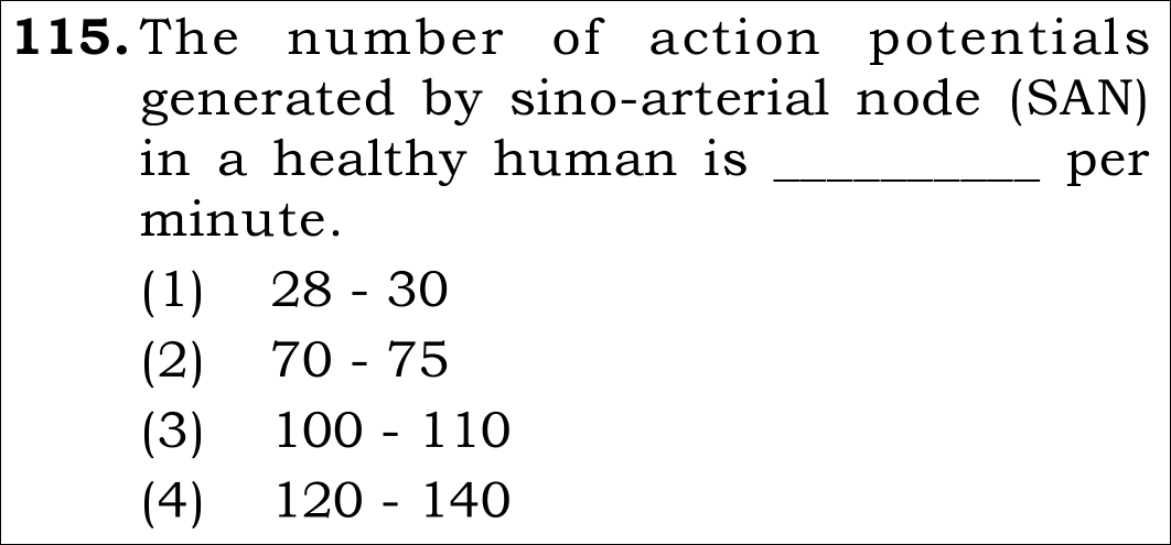
NEET 2026 · Q115 Easy
Official NEET 2026 Question 115 - Body Fluids and Circulation
1
2 ✓ Key
3
4
💡 Verified Solution & 30-Second Hack
✓
Verified Against NTA Final Answer Key
Option (2) : 70 - 75
📚 Body Fluids and Circulation Easy
📋 Given Parameters & Problem Setup
Anatomical Structure Sino-atrial node (SAN) / pacemaker of the human heart
Physiological Quantity Action potentials generated per minute under resting condition
⚡

The 30-Second Exam Hall Shortcut: ⚡ Resting Heart Rate Match

⏱️ Save ~40 seconds

SAN Action Potentials = Normal Heartbeat = 70–75 beats/min!

1 Recall that SAN sets the pace of the heart because it generates the highest frequency of spontaneous action potentials.
2 Normal resting heart rate is 72 beats/min, which reflects the SAN rate of 70–75 per minute.
3 Look at Option (2): 70 - 75.
4 Mark Option (2) in 3 seconds!
🛡️
Why You Can 100% Trust This Shortcut: NCERT Biology · Photosynthesis in Higher Plants (Historical Experiments: Joseph Priestley bell jar and mint plant experiment).
📐

Step-by-Step Rigorous Workflow & Mathematical Proof

Step 1

Electrophysiology of Cardiac Pacemaker

A specialized cardiac musculature called nodal tissue is distributed in the heart. A patch of this tissue called the Sino-atrial node (SAN) is located in the upper right corner of the right atrium.

SAN=Pacemaker of the human heart\text{SAN} = \text{Pacemaker of the human heart}
  • • Nodal cells possess inherent autorhythmicity (spontaneous diastolic depolarization) without external neural innervation.
Step 2

Quantify Action Potential Generation Rate

Different parts of the conducting system can generate action potentials at different inherent frequencies: SAN generates 70–75 min⁻¹, AVN generates 40–60 min⁻¹, and Purkinje fibers generate 20–40 min⁻¹. Because SAN possesses the highest intrinsic rate, it commands heart contraction.

FrequencySAN=70−75 action potentials⋅min−1\text{Frequency}_{\text{SAN}} = 70 - 75 \text{ action potentials} \cdot \text{min}^{-1}
  • • Dictates the average resting human heart rate of ~72 beats per minute.
Step 3

Evaluate Options

Option (1) gives 28-30 (characteristic of severe heart block / slow Purkinje rhythm). Option (3) gives 100-110 (tachycardia). Option (4) gives 120-140 (strenuous exercise). The basal rate is 70-75.

Correct Value=70−75  ⟹  Option (2)\text{Correct Value} = \mathbf{70 - 75} \implies \text{Option (2)}
  • • Matches official NTA final answer key Option B / (2).
⭐

360° Option Elimination Engine Key Feature

Diagnose every choice: Know exactly why the right option works and why distractors fail.

4-Option Deep Scan
1 A
✗ Incorrect
Verdict & Scientific Reason:

Incorrect. Represents ectopic ventricular/Purkinje fiber intrinsic firing rate.

⚠️
Student Fallacy (Why candidates pick this trap): Confusing pacemaker rate with lower conducting system fallback rate.
2 B
✓ 100% Correct
Verdict & Scientific Reason:

100% accurate. Canonical NCERT rate of SAN autorhythmicity.

💡
Why this is airtight: None; standard direct recall.
3 C
✗ Incorrect
Verdict & Scientific Reason:

Incorrect. Represents sinus tachycardia, not normal resting generation.

⚠️
Student Fallacy (Why candidates pick this trap): Overestimating resting rate.
4 D
✗ Incorrect
Verdict & Scientific Reason:

Incorrect. Exceeds standard physiological baseline.

⚠️
Student Fallacy (Why candidates pick this trap): Confusing normal rate with systolic pressure or exertion pulse.
⚠️

NTA Trap Alert: Typo in Official Exam Paper

The NEET question paper misprinted "sino-atrial node" as "sino-arterial node". Students should never get distracted by obvious typographical errors in anatomy names.

🛡️ How to avoid the -1 mark: Recognize the acronym (SAN) and focus on the physiological concept tested.
📖

Core NEET Syllabus Standard

NEET Biology Core Syllabus · Body Fluids and Circulation Standard Benchmark
Core Scientific Principle Tested:

"The sino-atrial node (SAN) generates 70–75 action potentials per minute, establishing it as the primary pacemaker of the mammalian heart."
Syllabus Focus: Circulatory Pathways - Cardiac Cycle and Nodal Tissue
✓ Core NEET UG Syllabus Anchor Syllabus & Academic Standards ↗
SAN sets the pulse alive: exactly seventy to seventy-five!
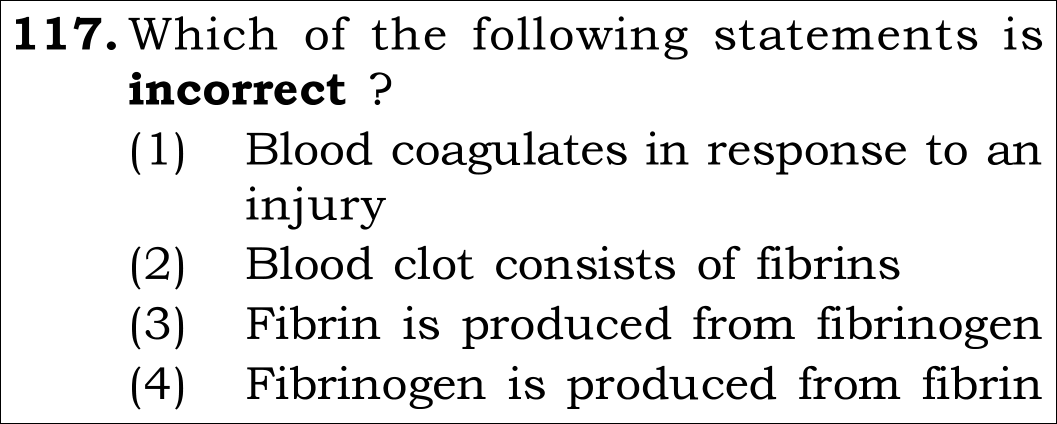
NEET 2026 · Q117 Easy
Official NEET 2026 Question 117 - Body Fluids and Circulation
1 Blood coagulates in response to an injury
2 Blood clot consists of fibrins
3 Fibrin is produced from fibrinogen
4 Fibrinogen is produced from fibrin ✓ Key
💡 Verified Solution & 30-Second Hack
✓
Verified Against NTA Final Answer Key
Option (4) : Fibrinogen is produced from fibrin
📚 Body Fluids and Circulation Easy
📋 Given Parameters & Problem Setup
Physiological Process Blood coagulation (hemostasis)
Key Components Thrombokinase, Prothrombin, Thrombin, Fibrinogen, Fibrin
⚡

The 30-Second Exam Hall Shortcut: ⚡ Clotting Precursor Direction Rule

⏱️ Save ~35 seconds

Thrombin turns inactive Fibrinogen INTO active Fibrin, NEVER the reverse!

1 Recall the final common pathway of coagulation:
2 Prothrombin -> Thrombin (via Thrombokinase + Ca²⁺).
3 Fibrinogen -> Fibrin (catalyzed by Thrombin).
4 Statement (3) correctly says "Fibrin is produced from fibrinogen".
5 Statement (4) falsely states "Fibrinogen is produced from fibrin". That is backwards!
6 The question asks for the INCORRECT statement -> Select Option (4) immediately!
🛡️
Why You Can 100% Trust This Shortcut: NCERT Body Fluids and Circulation verbatim: Fibrins are formed by the conversion of inactive fibrinogens in the plasma by the enzyme thrombin.
📐

Step-by-Step Rigorous Workflow & Mathematical Proof

Step 1

Understand the Blood Clotting Cascade

When a blood vessel suffers trauma or injury, platelets and injured tissues release thromboplastin, triggering an enzymatic cascade leading to the formation of thrombokinase (prothrombinase).

Prothrombin→Thrombokinase+Ca2+Thrombin\text{Prothrombin} \xrightarrow{\text{Thrombokinase} + \text{Ca}^{2+}} \text{Thrombin}
  • • Thrombokinase converts inactive plasma prothrombin into active thrombin.
Step 2

Fibrinogen to Fibrin Conversion

Thrombin then acts as an enzyme to cleave soluble inactive fibrinogen into insoluble fibrin monomers, which polymerize into long threads trapping blood cells to form the clot (coagulum).

Fibrinogen (soluble)→ThrombinFibrin (insoluble threads)\text{Fibrinogen (soluble)} \xrightarrow{\text{Thrombin}} \text{Fibrin (insoluble threads)}
  • • Therefore, Fibrin is derived from Fibrinogen, never Fibrinogen from Fibrin!
Step 3

Evaluate Each Statement

(1) Blood coagulates in response to injury: Correct physiological defense mechanism. (2) Blood clot consists of fibrins: Correct, fibrin meshwork forms the structural framework. (3) Fibrin is produced from fibrinogen: Correct. (4) Fibrinogen is produced from fibrin: Incorrect (inverted).

Incorrect Statement="Fibrinogen is produced from fibrin"  ⟹  Option (4)\text{Incorrect Statement} = \text{"Fibrinogen is produced from fibrin"} \implies \text{Option (4)}
  • • Matches official NTA final answer key Option D / (4).
⭐

360° Option Elimination Engine Key Feature

Diagnose every choice: Know exactly why the right option works and why distractors fail.

4-Option Deep Scan
1 A
✗ Incorrect
Verdict & Scientific Reason:

Factually correct statement, hence not the answer to this negative-stem question.

⚠️
Student Fallacy (Why candidates pick this trap): Failing to read the word "incorrect" in the question stem.
2 B
✗ Incorrect
Verdict & Scientific Reason:

Factually correct statement (coagulum is a mesh of fibrin threads trapping dead formed elements).

⚠️
Student Fallacy (Why candidates pick this trap): Confusing correct facts with incorrect choices.
3 C
✗ Incorrect
Verdict & Scientific Reason:

Factually correct biochemical reaction catalyzed by thrombin.

⚠️
Student Fallacy (Why candidates pick this trap): Not noticing the inversion between option 3 and option 4.
4 D
✓ 100% Correct
Verdict & Scientific Reason:

100% incorrect statement, making it the correct answer to the question. Fibrinogen is produced by hepatocytes in the liver, not from fibrin.

💡
Why this is airtight: None; carefully spotted the inverted precursor-product relationship.
⚠️

NTA Trap Alert: The Inversion Trap

Examiners place reciprocal statements (Statement 3 vs Statement 4) knowing that tired candidates may misread who converts into whom.

🛡️ How to avoid the -1 mark: The suffix "-ogen" always means precursor (inactive generator)! Fibrinogen GENERATES Fibrin; Pepsinogen GENERATES Pepsin.
📖

Core NEET Syllabus Standard

NEET Biology Core Syllabus · Body Fluids and Circulation Standard Benchmark
Core Scientific Principle Tested:

"In blood clotting, thrombin converts soluble fibrinogen into insoluble fibrin threads. Fibrinogen is never synthesized from fibrin."

Syllabus Focus: Coagulation of Blood
✓ Core NEET UG Syllabus Anchor Syllabus & Academic Standards ↗
-OGEN means the origin: Fibrinogen makes Fibrin, never reverse the spin!
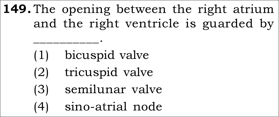
NEET 2026 · Q149 Easy
Official NEET 2026 Question 149 - Body Fluids and Circulation
1 bicuspid valve
2 tricuspid valve ✓ Key
3 semilunar valve
4 sino-atrial node
💡 Verified Solution & 30-Second Hack
✓
Verified Against NTA Final Answer Key
Option (2) : tricuspid valve
📚 Body Fluids and Circulation Easy
📋 Given Parameters & Problem Setup
Cardiac Aperture Opening between right atrium and right ventricle
Anatomical Structure Valve guarding this atrioventricular orifice
⚡

The 30-Second Exam Hall Shortcut: ⚡ Right = Tri; Left = Bi (MitrAL) Rule

⏱️ Save ~40 seconds

Right AV opening = TRICUSPID valve; Left AV opening = BICUSPID (Mitral) valve!

1 Recall anatomical valve rule:
2 Right atrioventricular orifice = guarded by 3 muscular flaps -> TRICUSPID valve.
3 Left atrioventricular orifice = guarded by 2 muscular flaps -> BICUSPID (Mitral) valve.
4 Semilunar valves guard pulmonary artery and aorta openings.
5 The question specifies "between right atrium and right ventricle".
6 Select Option (2) Tricuspid valve in 3 seconds!
🛡️
Why You Can 100% Trust This Shortcut: NCERT Biology · Body Fluids and Circulation (Cardiac Cycle: Stroke volume x Heart rate = Cardiac output of ~5 L/min).
📐

Step-by-Step Rigorous Workflow & Mathematical Proof

Step 1

Anatomy of Human Heart Valves

The human heart possesses four chambers: two atria and two ventricles. To ensure unidirectional blood flow and prevent backflow during ventricular systole, the atrioventricular apertures are guarded by specialized fibrous valves.

AV Valves={Right Atrium - Right Ventricle:Tricuspid Valve (Three flaps/cusps)Left Atrium - Left Ventricle:Bicuspid / Mitral Valve (Two flaps/cusps)\text{AV Valves} = \begin{cases} \mathbf{Right\text{ Atrium - Right Ventricle}} &: \mathbf{Tricuspid\text{ Valve (Three flaps/cusps)}} \\ \text{Left Atrium - Left Ventricle} &: \text{Bicuspid / Mitral Valve (Two flaps/cusps)} \end{cases}
  • • NCERT verbatim: "The opening between the right atrium and the right ventricle is guarded by a valve formed of three muscular flaps or cusps, the tricuspid valve."
Step 2

Evaluate Other Named Structures

Bicuspid (mitral) valve guards the left atrioventricular aperture. Semilunar valves guard the openings of the right ventricle into the pulmonary artery and left ventricle into the systemic aorta. The sino-atrial node (SAN) is the neuromuscular pacemaker patch, not a mechanical valve.

Right AV Valve=Tricuspid Valve  ⟹  Option (2)\text{Right AV Valve} = \mathbf{Tricuspid\text{ Valve}} \implies \text{Option (2)}
  • • Option (2) is the solely accurate response.
Step 3

Conclusion

The tricuspid valve guards the right atrioventricular aperture.

Correct Valve=Option (2)\text{Correct Valve} = \text{Option (2)}
  • • Matches official NTA final answer key Option B / (2).
⭐

360° Option Elimination Engine Key Feature

Diagnose every choice: Know exactly why the right option works and why distractors fail.

4-Option Deep Scan
1 A
✗ Incorrect
Verdict & Scientific Reason:

Incorrect. Bicuspid (mitral) valve guards the opening between the left atrium and left ventricle.

⚠️
Student Fallacy (Why candidates pick this trap): Swapping right and left cardiac valves.
2 B
✓ 100% Correct
Verdict & Scientific Reason:

100% accurate. The right atrioventricular opening is guarded by the tricuspid valve.

💡
Why this is airtight: None; standard direct anatomical recall.
3 C
✗ Incorrect
Verdict & Scientific Reason:

Incorrect. Semilunar valves guard the pulmonary and aortic outflow tract exits, not the atrioventricular orifices.

⚠️
Student Fallacy (Why candidates pick this trap): Confusing arterial outflow valves with atrioventricular valves.
4 D
✗ Incorrect
Verdict & Scientific Reason:

Incorrect. SAN is the electrical pacemaker tissue located in the right atrial wall, not a valve.

⚠️
Student Fallacy (Why candidates pick this trap): Confusing conducting nodal tissue with heart valves.
⚠️

NTA Trap Alert: Bicuspid vs Tricuspid Lateralization

Under exam pressure, students frequently forget whether Tricuspid is on the right or the left side of the heart.

🛡️ How to avoid the -1 mark: Remember: Try to be Right (TRIcuspid = RIGHT)!
📖

Core NEET Syllabus Standard

NEET Biology Core Syllabus · Body Fluids and Circulation Standard Benchmark
Core Scientific Principle Tested:

"The right atrioventricular aperture is guarded by the tricuspid valve; the left atrioventricular aperture is guarded by the bicuspid (mitral) valve."

Syllabus Focus: Circulatory Pathways - Human Circulatory System Anatomy
✓ Core NEET UG Syllabus Anchor Syllabus & Academic Standards ↗
TRI to be RIGHT: TRICUSPID is on the RIGHT; BI is on the LEFT!
NEET 2025 · Q112 Easy
Official NEET 2025 Question 112 - Body Fluids and Circulation
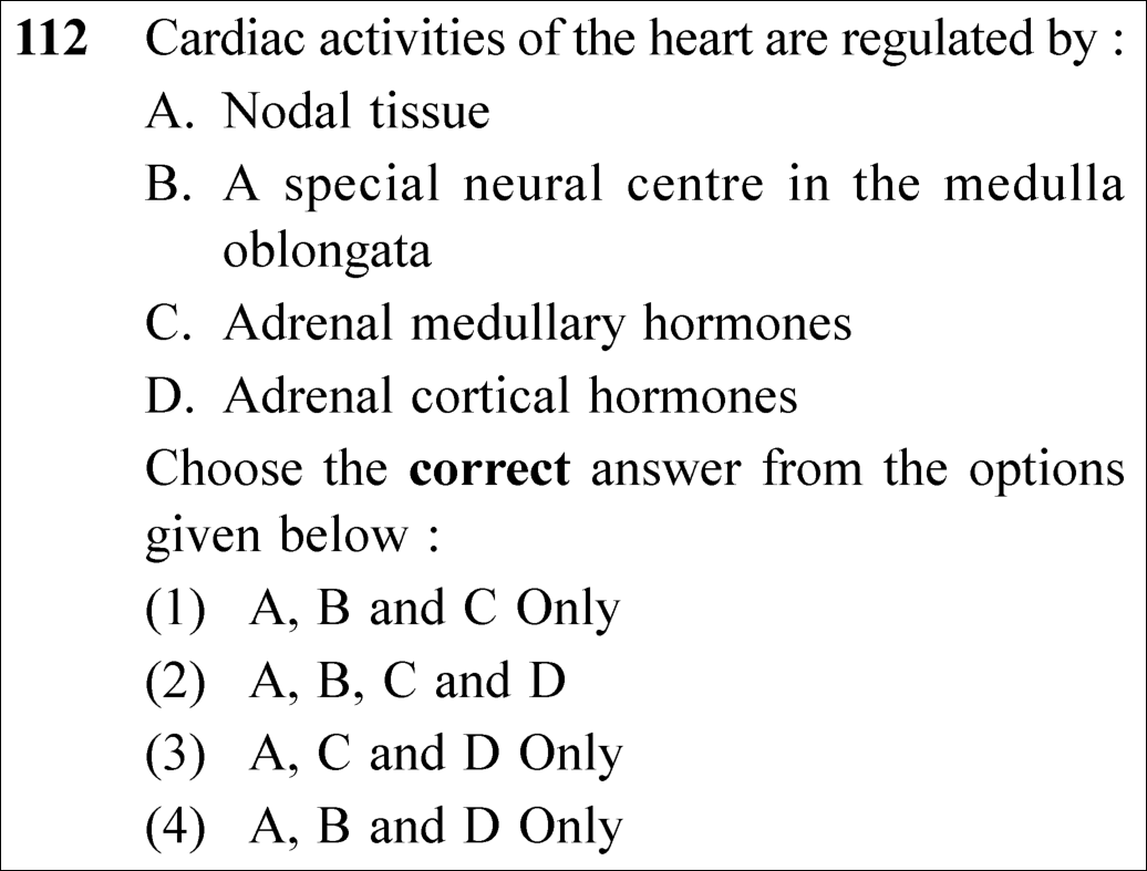
1 A, B and C Only ✓ Key
2 A,B, C and D
3 A, C and D Only
4 A, B and D Only
💡 Verified Solution & 30-Second Hack
✓
Verified Against NTA Final Answer Key
Option (1) : A, B and C Only
📚 Body Fluids and Circulation Easy
📋 Given Parameters & Problem Setup
Regulatory Mechanism A Nodal tissue
Regulatory Mechanism B A special neural centre in the medulla oblongata
Regulatory Mechanism C Adrenal medullary hormones
Regulatory Mechanism D Adrenal cortical hormones
⚡

The 30-Second Exam Hall Shortcut: ⚡ Cardiac Regulation Triad Fingerprint

⏱️ Save ~25 seconds

Heart rate regulated by: 1. Nodal tissue (SA node, auto-regulation myogenic), 2. Medulla oblongata (ANS sympathetic/parasympathetic), 3. Adrenal MEDULLA (epinephrine/norepinephrine)! Cortex is NOT in cardiac regulation triad!

1 Recall NCERT Section "Regulation of Cardiac Activity":
2 - Statement A: Nodal tissue auto-regulates heart intrinsically (myogenic). [CORRECT]
3 - Statement B: Special neural centre in medulla oblongata regulates heart through ANS. [CORRECT]
4 - Statement C: Adrenal medullary hormones (adrenaline/noradrenaline) increase heart rate and cardiac output. [CORRECT]
5 - Statement D: Adrenal cortical hormones (cortisol/aldosterone) regulate water-electrolyte balance and carbohydrates, not direct cardiac regulation. [INCORRECT]
6 A, B, and C Only are correct!
7 Select Option (1) immediately!
🛡️
Why You Can 100% Trust This Shortcut: Electrocardiogram (ECG) Deflections (NCERT Biology · Body Fluids and Circulation: P-wave represents atrial depolarisation).
📐

Step-by-Step Rigorous Workflow & Mathematical Proof

Step 1

Intrinsic (Auto-regulatory) Control by Nodal Musculature

Normal activities of the human heart are regulated intrinsically, i.e., auto-regulated by specialized cardiac muscle tissues called nodal tissue (Sinoatrial node, Atrioventricular node, bundle of His, Purkinje fibers). Hence, human heart is termed myogenic. Thus, Statement A is correct.

Step 2

Extrinsic Neural Regulation via Medulla Oblongata

A special neural center in the medulla oblongata moderates cardiac function through the autonomic nervous system (ANS):

  • • Sympathetic neural signals increase the rate of heartbeat, the strength of ventricular contraction, and thereby the cardiac output.
  • • Parasympathetic neural signals (via vagus nerve) decrease the rate of heartbeat, speed of action potential conduction, and cardiac output.
  • • Thus, Statement B is correct.
Step 3

Endocrine Regulation: Adrenal Medulla vs Cortex

Hormonal regulation of heart rate:

  • • Adrenal medullary hormones (epinephrine and norepinephrine / catecholamines) act during emergency / fight-or-flight states to significantly increase the rate of heartbeat, stroke volume, and cardiac output. Thus, Statement C is correct.
  • • Adrenal cortical hormones (glucocorticoids like cortisol and mineralocorticoids like aldosterone) are involved in carbohydrate metabolism, anti-inflammatory responses, and electrolyte balance (Na⁺/water retention in renal tubules), not direct cardiac pacing. Thus, Statement D is incorrect.
Step 4

Conclusion

Only A, B, and C are true regulators of cardiac activities according to the canonical NCERT text.

Regulation=A (Nodal)+B (Medulla)+C (Adrenal Medullary)\text{Regulation} = \text{A (Nodal)} + \text{B (Medulla)} + \text{C (Adrenal Medullary)}
⭐

360° Option Elimination Engine Key Feature

Diagnose every choice: Know exactly why the right option works and why distractors fail.

4-Option Deep Scan
1 A
✓ 100% Correct
Verdict & Scientific Reason:

100% correct. Matches the verbatim NCERT triad of cardiac regulation.

💡
Why this is airtight: None; precise physiological recall.
2 B
✗ Incorrect
Verdict & Scientific Reason:

Incorrect. Includes adrenal cortical hormones (D), which do not directly regulate cardiac activity.

⚠️
Student Fallacy (Why candidates pick this trap): Failing to distinguish adrenal medulla from adrenal cortex.
3 C
✗ Incorrect
Verdict & Scientific Reason:

Incorrect. Omits the critical neural center in the medulla oblongata (B) and improperly includes D.

⚠️
Student Fallacy (Why candidates pick this trap): Missing neural control of the heart.
4 D
✗ Incorrect
Verdict & Scientific Reason:

Incorrect. Omits adrenal medullary hormones (C) and includes cortical hormones (D).

⚠️
Student Fallacy (Why candidates pick this trap): Swapping medullary and cortical secretions.
⚠️

NTA Trap Alert: Adrenal Medullary vs Adrenal Cortical Hormones

The adrenal MEDULLA secretes adrenaline (epinephrine) which directly accelerates cardiac output. The adrenal CORTEX secretes steroids (aldosterone, cortisol). Never confuse medullary hormones with cortical hormones!

🛡️ How to avoid the -1 mark: Medulla = Adrenaline = Heart beat booster. Cortex = Steroids (sugar/salt balance).
📖

Core NEET Syllabus Standard

NEET Biology Core Syllabus · Body Fluids and Circulation Standard Benchmark
Core Scientific Principle Tested:

"Cardiac activities are regulated intrinsically by nodal tissue, neurally by medulla oblongata via ANS, and hormonally by adrenal medullary hormones."

Syllabus Focus: Regulation of Cardiac Activity
✓ Core NEET UG Syllabus Anchor Syllabus & Academic Standards ↗
N-M-M: Nodal tissue, Medulla oblongata, Medullary adrenal hormones!
NEET 2024 · Q176 Easy
Official NEET 2024 Question 176 - Body Fluids and Circulation
1
2
3 ✓ Key
4
💡 Verified Solution & 30-Second Hack
✓
Verified Against NTA Final Answer Key
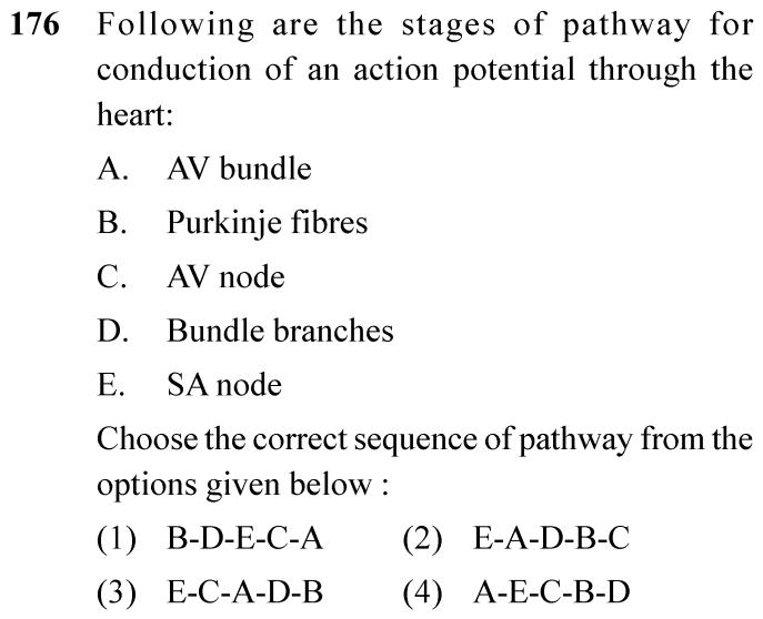
Option (3) : E-C-A-D-B
📚 Body Fluids and Circulation Easy
📋 Given Parameters & Problem Setup
Event Conduction of action potential through the human heart
Components A: AV bundle; B: Purkinje fibres; C: AV node; D: Bundle branches; E: SA node
⚡

The 30-Second Exam Hall Shortcut: ⚡ Cardiac Conduction Pathway Sequence

⏱️ Save ~25 seconds

SA node (Pacemaker E) -> AV node (C) -> AV bundle of His (A) -> Bundle branches (D) -> Purkinje fibres (B)! Sequence: E -> C -> A -> D -> B!

1 Recall the specialized nodal tissue sequence:
2 Sino-atrial node (SA node) initiates the impulse at upper right corner of right atrium (E).
3 Atrio-ventricular node (AV node) receives impulse at lower left corner of right atrium (C).
4 AV bundle (Bundle of His) carries impulse through interventricular septum (A).
5 Right and left bundle branches divide into ventricular walls (D).
6 Purkinje fibres transmit impulse throughout ventricular musculature (B).
7 Correct sequence: E -> C -> A -> D -> B -> Option (3) / C.
🛡️
Why You Can 100% Trust This Shortcut: NCERT Biology · Body Fluids and Circulation (Conducting System of the Human Heart)
📐

Step-by-Step Rigorous Workflow & Mathematical Proof

Step 1

Impulse Initiation at the Pacemaker

The sino-atrial node (SAN) can generate the maximum number of action potentials, i.e., 70-75 min⁻¹, and is responsible for initiating and maintaining the rhythmic contractile activity of the heart. Therefore, it is called the pacemaker (Stage E).

Step 2

Nodal Transmission to Ventricles

The action potential spreads from the SAN to the atrio-ventricular node (AVN) situated in the lower left corner of the right atrium close to the atrio-ventricular septum (Stage C). From the AVN, an atrioventricular bundle (AV bundle / bundle of His) continues that divides into a right and left bundle (Stages A and D).

Step 3

Ventricular Excitation via Purkinje Fibres

These branches give rise to minute fibres throughout the ventricular musculature of the respective sides called Purkinje fibres (Stage B), causing simultaneous ventricular contraction.

⭐

360° Option Elimination Engine Key Feature

Diagnose every choice: Know exactly why the right option works and why distractors fail.

4-Option Deep Scan
1 (1)
✗ Incorrect
Verdict & Scientific Reason:

Starts at Purkinje fibres instead of the pacemaker.

⚠️
Student Fallacy (Why candidates pick this trap): Starts at Purkinje fibres instead of the pacemaker.
2 (2)
✗ Incorrect
Verdict & Scientific Reason:

Places AV node at the end instead of directly after SA node.

⚠️
Student Fallacy (Why candidates pick this trap): Places AV node at the end instead of directly after SA node.
3 (3)
✓ 100% Correct
Verdict & Scientific Reason:

Accurately traces anatomical conduction: SA node -> AV node -> AV bundle -> Bundle branches -> Purkinje fibres.

💡
Why this is airtight: None.
4 (4)
✗ Incorrect
Verdict & Scientific Reason:

Places AV bundle before SA node.

⚠️
Student Fallacy (Why candidates pick this trap): Places AV bundle before SA node.
⚠️

NTA Trap Alert: AV Bundle vs AV Node Order

Swapping AV node and AV bundle in the sequence.

🛡️ How to avoid the -1 mark: Node comes first (receives atrial impulse), then the Bundle conducts down the septum.
📖

Core NEET Syllabus Standard

Biology Core Zoology · Body Fluids and Circulation Standard Benchmark
Core Scientific Principle Tested:

"The SAN can generate the maximum number of action potentials... An atrio-ventricular bundle continues from the AVN which passes through the atrio-ventricular septa to emerge on the top of the interventricular septum and immediately divides into a right and left bundle. These branches give rise to minute fibres called Purkinje fibres."

Syllabus Focus: Nodal Conduction Pathway of the Heart Action Potential
✓ Core NEET UG Syllabus Anchor Syllabus & Academic Standards ↗
NEET 2024 · Q190 Easy
Official NEET 2024 Question 190 - Body Fluids and Circulation
1
2
3
4 ✓ Key
💡 Verified Solution & 30-Second Hack
✓
Verified Against NTA Final Answer Key
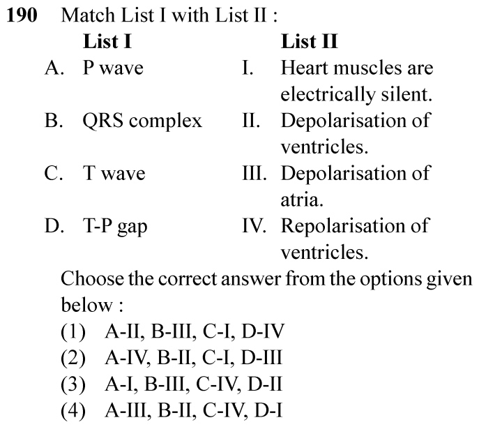
Option (4) : A-III, B-II, C-IV, D-I
📚 Body Fluids and Circulation Easy
📋 Given Parameters & Problem Setup
List I (ECG Deflections) A: P wave; B: QRS complex; C: T wave; D: T-P gap
List II (Electrophysiological Events) I: Heart muscles are electrically silent; II: Depolarisation of ventricles; III: Depolarisation of atria; IV: Repolarisation of ventricles
⚡

The 30-Second Exam Hall Shortcut: ⚡ ECG Wave Diagnostic Match

⏱️ Save ~25 seconds

P wave = Atrial depolarisation (III); QRS = Ventricular depolarisation (II); T wave = Ventricular repolarisation (IV); T-P gap = Electrical silence / diastole (I)! Match: A-III, B-II, C-IV, D-I!

1 A. P wave: Electrical excitation / Depolarisation of the atria (III).
2 B. QRS complex: Depolarisation of the ventricles initiating ventricular systole (II).
3 C. T wave: Return of ventricles from excited to normal state / Repolarisation of ventricles (IV).
4 D. T-P gap: Period between ventricular repolarisation and next atrial depolarisation during which the heart is in joint diastole / electrically silent (I).
5 Matching order: A-III, B-II, C-IV, D-I -> Option (4) / D.
🛡️
Why You Can 100% Trust This Shortcut: NCERT Biology · Body Fluids and Circulation (Electrocardiograph and ECG Waves)
📐

Step-by-Step Rigorous Workflow & Mathematical Proof

Step 1

Review Classical Electrocardiogram (ECG) Waves

Each peak in the ECG is identified with a letter from P to T that corresponds to a specific electrical activity of the heart: - The P-wave represents the electrical excitation (or depolarisation) of the atria, which leads to the contraction of both the atria (A -> III). - The QRS complex represents the depolarisation of the ventricles, which initiates the ventricular contraction (B -> II). - The T-wave represents the return of the ventricles from excited to normal state (repolarisation) (C -> IV).

Step 2

Define the T-P Interval / Gap

The interval between the end of the T wave and the beginning of the next P wave (T-P segment) represents the period of complete cardiac relaxation (joint diastole) during which ventricles and atria are relaxing and the heart is electrically silent (D -> I).

Step 3

Select Matching Combination

A-III, B-II, C-IV, D-I corresponds to Option (4).

⭐

360° Option Elimination Engine Key Feature

Diagnose every choice: Know exactly why the right option works and why distractors fail.

4-Option Deep Scan
1 (1)
✗ Incorrect
Verdict & Scientific Reason:

Mismatches P wave with ventricular depolarisation.

⚠️
Student Fallacy (Why candidates pick this trap): Mismatches P wave with ventricular depolarisation.
2 (2)
✗ Incorrect
Verdict & Scientific Reason:

Mismatches P wave with ventricular repolarisation.

⚠️
Student Fallacy (Why candidates pick this trap): Mismatches P wave with ventricular repolarisation.
3 (3)
✗ Incorrect
Verdict & Scientific Reason:

Mismatches P wave with electrical silence.

⚠️
Student Fallacy (Why candidates pick this trap): Mismatches P wave with electrical silence.
4 (4)
✓ 100% Correct
Verdict & Scientific Reason:

Accurately pairs all four ECG features with their definitive electrophysiological events.

💡
Why this is airtight: None.
⚠️

NTA Trap Alert: T Wave vs QRS Repolarisation

Confusing atrial repolarisation with the T wave.

🛡️ How to avoid the -1 mark: Atrial repolarisation is buried inside the large QRS complex; the T wave specifically represents VENTRICULAR repolarisation.
📖

Core NEET Syllabus Standard

Biology Core Zoology · Body Fluids and Circulation Standard Benchmark
Core Scientific Principle Tested:

"The P-wave represents the electrical excitation (or depolarisation) of the atria... The QRS complex represents the depolarisation of the ventricles... The T-wave represents the return of the ventricles from excited to normal state (repolarisation)."

Syllabus Focus: Standard ECG Deflection Waves and Corresponding Cardiac Phases
✓ Core NEET UG Syllabus Anchor Syllabus & Academic Standards ↗
NEET 2023 · Q153 Easy
Official NEET 2023 Question 153 - Body Fluids and Circulation
1
2 ✓ Key
3
4
💡 Verified Solution & 30-Second Hack
✓
Verified Against NTA Final Answer Key
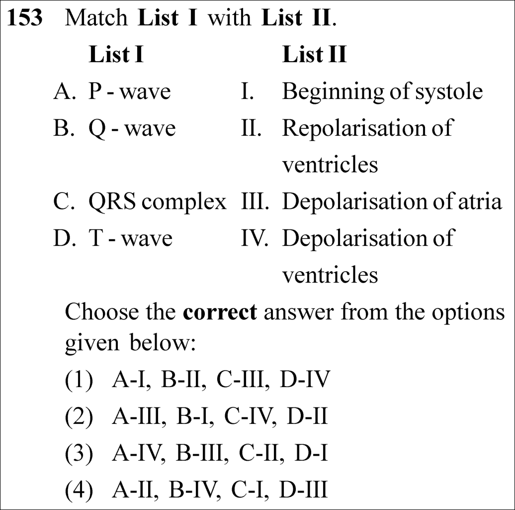
Option (2) : A-III, B-I, C-IV, D-II
📚 Body Fluids and Circulation Easy
📋 Given Parameters & Problem Setup
ECG Waves P-wave, Q-wave, QRS complex, T-wave
Cardiac Cycle Events Depolarisation of atria, Beginning of systole, Depolarisation of ventricles, Repolarisation of ventricles
⚡

The 30-Second Exam Hall Shortcut: ⚡ Standard ECG Wave Decoder

⏱️ Save ~25 seconds

Match standard ECG waves: P = Atrial depolarisation (III); QRS = Ventricular depolarisation (IV); T = Ventricular repolarisation (II); Q-wave = Marks beginning of ventricular systole (I)! Match Option (2)!

1 P-wave: Electrical excitation / depolarisation of atria -> A-III.
2 QRS complex: Depolarisation of ventricles (initiates ventricular contraction) -> C-IV.
3 Q-wave: Marks the beginning of the ventricular systole -> B-I.
4 T-wave: Repolarisation of ventricles (return to resting state) -> D-II.
5 Match combination: A-III, B-I, C-IV, D-II -> Option (2).
🛡️
Why You Can 100% Trust This Shortcut: Canonical NCERT Human Physiology electrocardiography electrophysiological waves.
📐

Step-by-Step Rigorous Workflow & Mathematical Proof

Step 1

Electrophysiological Basis of ECG Waves

A standard electrocardiogram represents the sequential propagation of action potentials across cardiac chambers:

A. P-wave:Depolarisation of the atria (leads to atrial contraction)  ⟹  IIIB. Q-wave:Marks the beginning of ventricular systole  ⟹  IC. QRS complex:Depolarisation of the ventricles (initiates ventricular systole)  ⟹  IVD. T-wave:Repolarisation of the ventricles (end of ventricular systole)  ⟹  II\begin{aligned} \text{\textbf{A. P-wave:}} & \quad \text{Depolarisation of the atria (leads to atrial contraction)} \implies \mathbf{\text{III}} \\[4pt] \text{\textbf{B. Q-wave:}} & \quad \text{Marks the beginning of ventricular systole} \implies \mathbf{\text{I}} \\[4pt] \text{\textbf{C. QRS complex:}} & \quad \text{Depolarisation of the ventricles (initiates ventricular systole)} \implies \mathbf{\text{IV}} \\[4pt] \text{\textbf{D. T-wave:}} & \quad \text{Repolarisation of the ventricles (end of ventricular systole)} \implies \mathbf{\text{II}} \end{aligned}
Step 2

Verbatim NCERT Reference

NCERT states under Electrocardiograph (ECG): - "The P-wave represents the electrical excitation (or depolarisation) of the atria..." - "The QRS complex represents the depolarisation of the ventricles..." - "The contraction starts shortly after Q and marks the beginning of the systole." - "The T-wave represents the return of the ventricles from excited to normal state (repolarisation)."

Step 3

Option Elimination

Matching A-III immediately isolates Option (2), making this solvable within seconds.

⭐

360° Option Elimination Engine Key Feature

Diagnose every choice: Know exactly why the right option works and why distractors fail.

4-Option Deep Scan
1 1
✗ Incorrect
Verdict & Scientific Reason:

Mismatches P-wave with beginning of systole.

⚠️
Student Fallacy (Why candidates pick this trap): Confusing atrial electrical activity with mechanical systole.
2 2
✓ 100% Correct
Verdict & Scientific Reason:

Flawless 1-to-1 match with the canonical NCERT electrocardiography definitions.

💡
Why this is airtight: None; textbook verbatim match.
3 3
✗ Incorrect
Verdict & Scientific Reason:

Mismatches P-wave with ventricular depolarisation.

⚠️
Student Fallacy (Why candidates pick this trap): Inverting atrial and ventricular waves.
4 4
✗ Incorrect
Verdict & Scientific Reason:

Mismatches P-wave with repolarisation.

⚠️
Student Fallacy (Why candidates pick this trap): Confusing depolarisation with repolarisation.
⚠️

NTA Trap Alert: Q-wave vs. QRS Complex Clinical Trap

Students often overlook the specific isolated role of the Q-wave: while the entire QRS complex represents ventricular depolarisation, the Q-wave specifically marks the onset/beginning of ventricular systole.

🛡️ How to avoid the -1 mark: Remember: QRS = Ventricular depolarisation. Q-wave alone = Beginning of systole.
📖

Core NEET Syllabus Standard

NCERT Biology · Body Fluids and Circulation Standard Benchmark
Core Scientific Principle Tested:

"P-wave is atrial depolarisation; Q-wave marks the beginning of systole; QRS complex is ventricular depolarisation; T-wave is ventricular repolarisation."

Syllabus Focus: Electrocardiograph (ECG)
✓ Core NEET UG Syllabus Anchor Syllabus & Academic Standards ↗
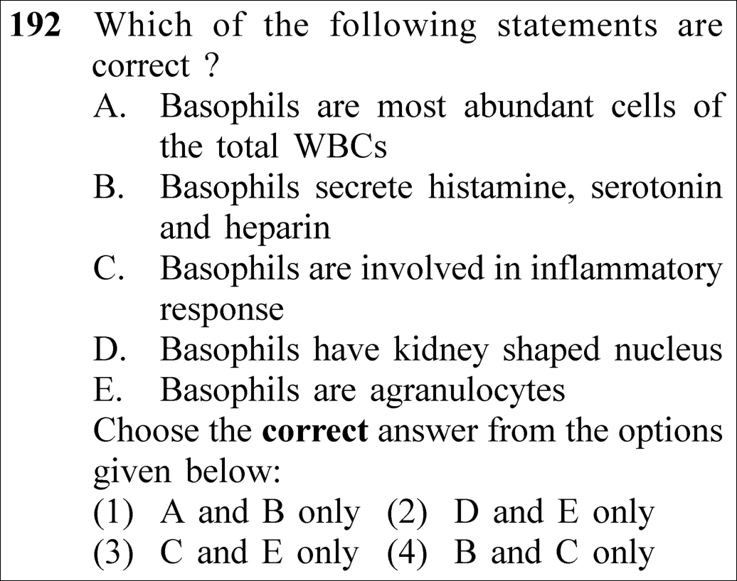
NEET 2023 · Q192 Easy
Official NEET 2023 Question 192 - Body Fluids and Circulation
1 A and B only
2 D and E only
3 C and E only
4 B and C only ✓ Key
💡 Verified Solution & 30-Second Hack
✓
Verified Against NTA Final Answer Key
Option (4) : B and C only
📚 Body Fluids and Circulation Easy
📋 Given Parameters & Problem Setup
Cell Type Basophils (Granulocytic White Blood Cells)
Statements 5 statements (A-E) on abundance, secretions, inflammation, nucleus, and granule status
⚡

The 30-Second Exam Hall Shortcut: ⚡ Basophil Fact Checker: Least Abundant Granulocyte!

⏱️ Save ~25 seconds

Basophils are the LEAST abundant WBCs (0.5–1%, ruling out A). They are GRANULOCYTES (ruling out E) with S-shaped/bilobed nuclei (monocytes have kidney-shaped, ruling out D). They secrete histamine, serotonin, heparin (B) and mediate inflammation (C)! Match Option (4) B and C only!

1 Statement A: Basophils are most abundant -> False (Neutrophils are most abundant 60–65%; basophils are least abundant 0.5–1%).
2 Statement B: Secrete histamine, serotonin, heparin -> True.
3 Statement C: Involved in inflammatory responses -> True.
4 Statement D: Kidney-shaped nucleus -> False (Kidney-shaped is monocyte; basophil has S-shaped/irregular 2–3 lobed nucleus).
5 Statement E: Basophils are agranulocytes -> False (They are granulocytes).
6 B and C only -> Select Option (4).
🛡️
Why You Can 100% Trust This Shortcut: Canonical NCERT Human Physiology formed elements and leukocyte profiles.
📐

Step-by-Step Rigorous Workflow & Mathematical Proof

Step 1

Evaluation of Statements B and C (Valid Statements)

Basophils are circulating granulocytes containing coarse basophilic granules (staining with basic dyes like methylene blue). Their granules release potent inflammatory vasoactive mediators:

Basophils⟶{Histamine(Vasodilator)Serotonin(Vasoconstrictor)Heparin(Anticoagulant)  ⟹  Mediate Inflammatory Responses\text{\textbf{Basophils}} \longrightarrow \begin{cases} \mathbf{\text{Histamine}} & (\text{Vasodilator}) \\[2pt] \mathbf{\text{Serotonin}} & (\text{Vasoconstrictor}) \\[2pt] \mathbf{\text{Heparin}} & (\text{Anticoagulant}) \end{cases} \implies \mathbf{\text{Mediate Inflammatory Responses}}
Step 2

Evaluation of Statements A, D, and E (Invalid Statements)

Analyzing the errors in the false statements: - Statement A: Neutrophils are the most abundant (60−65%60-65\%). Basophils are the least abundant (0.5−1%0.5-1\%). - Statement D: Monocytes have a characteristic large kidney-shaped/bean-shaped nucleus. Basophils have an S-shaped or bilobed nucleus obscured by dark granules. - Statement E: Granulocytes include neutrophils, eosinophils, and basophils. Agranulocytes include lymphocytes and monocytes.

Step 3

Verbatim NCERT Reference

NCERT states verbatim: "Basophils are least (0.5-1 per cent) among them. Basophils secrete histamine, serotonin, heparin, etc., and are involved in inflammatory reactions."

⭐

360° Option Elimination Engine Key Feature

Diagnose every choice: Know exactly why the right option works and why distractors fail.

4-Option Deep Scan
1 1
✗ Incorrect
Verdict & Scientific Reason:

Includes false statement A (basophils are the least, not most, abundant WBCs).

⚠️
Student Fallacy (Why candidates pick this trap): Confusing basophils with neutrophils in relative abundance.
2 2
✗ Incorrect
Verdict & Scientific Reason:

Both D (kidney-shaped nucleus describes monocytes) and E (basophils are granulocytes) are false.

⚠️
Student Fallacy (Why candidates pick this trap): Confusing agranulocyte morphology with basophils.
3 3
✗ Incorrect
Verdict & Scientific Reason:

Includes false statement E.

⚠️
Student Fallacy (Why candidates pick this trap): Calling basophils agranulocytes.
4 4
✓ 100% Correct
Verdict & Scientific Reason:

Statements B and C are completely accurate textbook facts regarding basophil physiology.

💡
Why this is airtight: None; textbook verbatim precision.
⚠️

NTA Trap Alert: Monocyte vs. Basophil Nucleus Morphology Trap

Monocytes have a kidney/bean-shaped nucleus. Eosinophils have a bilobed spectacle-shaped nucleus. Neutrophils have multi-lobed (3-5 lobes) nucleus. Basophils have an S-shaped/irregular nucleus.

🛡️ How to avoid the -1 mark: Kidney-shaped = Monocyte. S-shaped with dark granules = Basophil.
📖

Core NEET Syllabus Standard

NCERT Biology · Body Fluids and Circulation Standard Benchmark
Core Scientific Principle Tested:

"Basophils secrete histamine, serotonin, heparin, and are involved in inflammatory reactions. They are the least abundant granulocytes."

Syllabus Focus: Formed Elements (Leukocytes)
✓ Core NEET UG Syllabus Anchor Syllabus & Academic Standards ↗
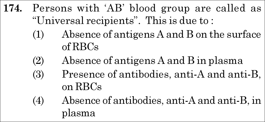
NEET 2021 · Q174 Easy
Official NEET 2021 Question 174 - Body Fluids and Circulation
1 Absence of antigens A and B on the surface of RBCs
2 Absence of antigens A and B in plasma
3 Presence of antibodies, anti-A and anti-B, on RBCs
4 Absence of antibodies, anti-A and anti-B, in plasma ✓ Key
💡 Verified Solution & 30-Second Hack
✓
Verified Against NTA Final Answer Key
Option (4) : Absence of antibodies, anti-A and anti-B, in plasma
📚 Body Fluids and Circulation Easy
📋 Given Parameters & Problem Setup
Phenotype AB Blood Group
Clinical Title Universal Recipient
Question Objective Identify the immunological basis for being a universal recipient
⚡

The 30-Second Exam Hall Shortcut: ⚡ "AB Group = Has A & B Antigens on RBCs, ZERO Antibodies in Plasma"

⏱️ Save ~20 seconds

Because AB blood individuals produce NEITHER anti-A NOR anti-B antibodies in their blood plasma, foreign transfused RBCs carrying A or B antigens will not be agglutinated or destroyed. Thus, absence of plasma antibodies enables universal reception!

1 Recall Landsteiner rule: Antigens reside on RBC membranes; Antibodies reside in blood plasma.
2 Blood group AB profile: - Surface Antigens: Both Antigen A and Antigen B present. - Plasma Antibodies: Neither anti-A nor anti-B (absent!).
3 Transfusion compatibility: Without recipient antibodies, donor RBCs are not attacked.
4 Match Option (4): "Absence of antibodies, anti-A and anti-B, in plasma".
🛡️
Why You Can 100% Trust This Shortcut: NCERT Biology · Body Fluids and Circulation (Blood Groups: Persons with AB group can accept blood from persons with AB as well as the other groups of blood. Therefore, such persons are called universal recipients because they have both A and B antigens on RBCs but no antibodies in plasma).
📐

Step-by-Step Rigorous Workflow & Mathematical Proof

Step 1

Immunological Profile of ABO Blood Groups

The ABO blood group system is governed by surface oligosaccharide antigens on red blood cell membranes and naturally occurring isoantibodies in blood plasma: - Group A: Antigen A on RBCs; anti-B antibody in plasma. - Group B: Antigen B on RBCs; anti-A antibody in plasma. - Group AB: Both Antigen A and Antigen B on RBCs; NEITHER anti-A nor anti-B antibodies in plasma. - Group O: No A or B antigens on RBCs; both anti-A and anti-B antibodies in plasma.

Group AB  ⟹  {RBC Surface:Antigens A and B PresentBlood Plasma:Anti-A and Anti-B Antibodies ABSENT\text{Group AB} \implies \begin{cases} \textbf{RBC Surface:} & \text{Antigens A and B Present} \\[3pt] \textbf{Blood Plasma:} & \text{Anti-A and Anti-B Antibodies } \mathbf{ABSENT} \end{cases}
Step 2

Mechanism of Universal Reception

When donor blood is transfused, the critical clinical danger is agglutination and haemolysis of the donor RBCs by antibodies present in the recipient's plasma. Because an individual with blood group AB has zero circulating anti-A and anti-B antibodies, transfused erythrocytes of type A, B, AB, or O encounter no opposing agglutinins, allowing safe transfusion without immune destruction.

⭐

360° Option Elimination Engine Key Feature

Diagnose every choice: Know exactly why the right option works and why distractors fail.

4-Option Deep Scan
1 1
✗ Incorrect
Verdict & Scientific Reason:

Incorrect. Absence of antigens on RBCs defines blood group O (universal donor), not blood group AB.

⚠️
Student Fallacy (Why candidates pick this trap): Confusing blood group O (no antigens on RBC) with blood group AB (no antibodies in plasma).
2 2
✗ Incorrect
Verdict & Scientific Reason:

Incorrect. Antigens are membrane-bound surface proteins/glycolipids on erythrocytes, not plasma solutes.

⚠️
Student Fallacy (Why candidates pick this trap): Confusing the anatomical compartments of antigens (RBC surface) and antibodies (plasma).
3 3
✗ Incorrect
Verdict & Scientific Reason:

Incorrect. Antibodies are soluble immunoglobulins in plasma, not surface receptors on RBCs.

⚠️
Student Fallacy (Why candidates pick this trap): Conflating antigens and antibodies.
4 4
✓ 100% Correct
Verdict & Scientific Reason:

Correct. Lacking both anti-A and anti-B antibodies in plasma prevents agglutination of any incoming donor red blood cells.

💡
Why this is airtight: None. Candidate accurately pairs the absence of antibodies in recipient plasma with universal recipient status.
⚠️

NTA Trap Alert: Antigen vs Antibody Compartment Inversion Trap

Options 1 and 2 swap antigens onto plasma or eliminate them from RBCs. Students rushing through the exam frequently read "absence of..." and pick Option 1, inadvertently selecting the feature of blood group O.

🛡️ How to avoid the -1 mark: Remember: Universal Donor (O) = Zero Antigens on RBC; Universal Recipient (AB) = Zero Antibodies in Plasma.
📖

Core NEET Syllabus Standard

NEET / NCERT Biology · Body Fluids and Circulation Standard Benchmark
Core Scientific Principle Tested:

"Persons with AB blood group are universal recipients because their plasma lacks both anti-A and anti-B antibodies."

Syllabus Focus: ABO Blood Grouping
✓ Core NEET UG Syllabus Anchor Syllabus & Academic Standards ↗
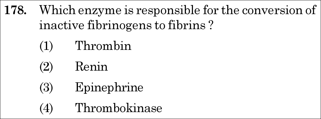
NEET 2021 · Q178 Easy
Official NEET 2021 Question 178 - Body Fluids and Circulation
1 Thrombin ✓ Key
2 Renin
3 Epinephrine
4 Thrombokinase
💡 Verified Solution & 30-Second Hack
✓
Verified Against NTA Final Answer Key
Option (1) : Thrombin
📚 Body Fluids and Circulation Easy
📋 Given Parameters & Problem Setup
Substrate Inactive soluble Fibrinogen
Product Insoluble Fibrin threads / clot network
Question Objective Identify the specific enzyme catalyzing this proteolytic cleavage
⚡

The 30-Second Exam Hall Shortcut: ⚡ "Cascade Rule: Thrombokinase activates Prothrombin → Thrombin cleaves Fibrinogen"

⏱️ Save ~20 seconds

Thrombokinase converts Prothrombin to Thrombin; then active Thrombin cleaves soluble Fibrinogen into insoluble Fibrin meshwork! Therefore, the direct enzyme converting fibrinogen to fibrin is Thrombin. Mark Option (1)!

1 Recall the 2-step terminal coagulation cascade: - Step 1: Prothrombin →Thrombokinase+Ca2+\xrightarrow{\text{Thrombokinase} + Ca^{2+}} Thrombin. - Step 2: Fibrinogen →Thrombin\xrightarrow{\textbf{Thrombin}} Fibrin.
2 Identify the direct catalyst of Step 2: Thrombin.
3 Avoid distractor: Thrombokinase acts upstream on prothrombin, not directly on fibrinogen.
4 Select Option (1).
🛡️
Why You Can 100% Trust This Shortcut: NCERT Biology · Body Fluids and Circulation (Coagulation of Blood: Fibrins are formed by the conversion of inactive fibrinogens in the plasma by the enzyme thrombin. Thrombins, in turn are formed from another inactive substance present in the plasma called prothrombin).
📐

Step-by-Step Rigorous Workflow & Mathematical Proof

Step 1

Enzymatic Cascade of Blood Coagulation

Coagulation is an amplified biological cascade where inactive proenzymes are sequentially converted to active serine proteases: 1. Injured tissues and activated platelets release thromboplastin, forming the enzyme complex Thrombokinase (prothrombinase). 2. In the presence of ionic calcium (Ca2+Ca^{2+}), thrombokinase catalyzes the proteolytic conversion of the plasma zymogen Prothrombin into active Thrombin. 3. Thrombin acts directly on soluble globular Fibrinogen (Factor I), cleaving fibrinopeptides A and B to yield insoluble fibrin monomers that polymerize and cross-link into a meshwork trapping blood cells to form the definitive clot.

Prothrombin→Ca2+ThrombokinaseThrombinInactive Fibrinogen→ThrombinInsoluble Fibrin Clot\begin{aligned} \text{Prothrombin} &\xrightarrow[Ca^{2+}]{\text{Thrombokinase}} \mathbf{Thrombin} \\[6pt] \text{Inactive Fibrinogen} &\xrightarrow{\mathbf{Thrombin}} \mathbf{Insoluble\,Fibrin\,Clot} \end{aligned}
Step 2

Elimination of Non-Coagulation Options

- Renin: An aspartic protease secreted by juxtaglomerular cells of the kidney that converts angiotensinogen into angiotensin I (RAAS pathway). - Epinephrine (Adrenaline): An amino acid derivative hormone secreted by the adrenal medulla during fight-or-flight emergencies. - Thrombokinase: Activates prothrombin to thrombin; it does not directly convert fibrinogen to fibrin.

⭐

360° Option Elimination Engine Key Feature

Diagnose every choice: Know exactly why the right option works and why distractors fail.

4-Option Deep Scan
1 1
✓ 100% Correct
Verdict & Scientific Reason:

Correct. Thrombin is the specific serine protease that cleaves fibrinogen into insoluble fibrin.

💡
Why this is airtight: None. Candidate remembers the exact enzymatic substrate of thrombin.
2 2
✗ Incorrect
Verdict & Scientific Reason:

Incorrect. Renin is a renal hormone involved in blood pressure regulation via the RAAS axis.

⚠️
Student Fallacy (Why candidates pick this trap): Confusing renin with coagulating enzymes due to rough phonetic similarity with rennin.
3 3
✗ Incorrect
Verdict & Scientific Reason:

Incorrect. Epinephrine is an emergency catecholamine hormone, not a clotting protease.

⚠️
Student Fallacy (Why candidates pick this trap): Mistaking sympathetic vasoconstrictors for biochemical clotting catalysts.
4 4
✗ Incorrect
Verdict & Scientific Reason:

Incorrect. Thrombokinase converts prothrombin into thrombin, which is the immediate upstream reaction.

⚠️
Student Fallacy (Why candidates pick this trap): Failing to distinguish between the initiator enzyme (thrombokinase) and the terminal effector enzyme (thrombin).
⚠️

NTA Trap Alert: Thrombokinase vs Thrombin Cascade Step Trap

Candidates know that thrombokinase and thrombin are both involved in clotting. The trap lies in confusing which enzyme acts on prothrombin (thrombokinase) versus which enzyme acts directly on fibrinogen (thrombin).

🛡️ How to avoid the -1 mark: Remember: Thrombokinase makes Thrombin; Thrombin makes Fibrin.
📖

Core NEET Syllabus Standard

NEET / NCERT Biology · Body Fluids and Circulation Standard Benchmark
Core Scientific Principle Tested:

"Thrombin converts inactive fibrinogen into insoluble fibrin threads."

Syllabus Focus: Coagulation of Blood
✓ Core NEET UG Syllabus Anchor Syllabus & Academic Standards ↗
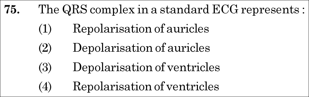
NEET 2020 · Q75 Easy
Official NEET 2020 Question 75 - Body Fluids and Circulation
1 Repolarisation of auricles
2 Depolarisation of auricles
3 Depolarisation of ventricles ✓ Key
4 Repolarisation of ventricles
💡 Verified Solution & 30-Second Hack
✓
Verified Against NTA Final Answer Key
Option (3) : Depolarisation of ventricles
📚 Body Fluids and Circulation Easy
📋 Given Parameters & Problem Setup
Diagnostic Waveform QRS complex in a standard ECG
Question Objective Identify which cardiac electrophysiological event is represented
⚡

The 30-Second Exam Hall Shortcut: ⚡ "P = Atrial Depol; QRS = Ventricular Depol; T = Ventricular Repol!"

⏱️ 20 seconds

P wave represents depolarisation of atria. QRS complex represents DEPOLARISATION OF VENTRICLES (triggering ventricular contraction/systole). T wave represents repolarisation of ventricles. Mark Depolarisation of ventricles (Option 3) in 5 seconds!

1 P-wave = Depolarisation of the atria.
2 QRS complex = Depolarisation of the ventricles.
3 T-wave = Repolarisation of the ventricles.
4 Select Option (3) Depolarisation of ventricles.
🛡️
Why You Can 100% Trust This Shortcut: NCERT Biology · Human Physiology: Body Fluids and Circulation (Electrocardiograph: ECG)
📐

Step-by-Step Rigorous Workflow & Mathematical Proof

Step 1

Review the Components of a Standard ECG

A standard ECG tracing records the electrical changes propagating through the myocardium during a cardiac cycle, consisting of a P wave, a QRS complex, and a T wave.

Step 2

Analyze the QRS Complex

The QRS complex represents the electrical depolarisation of the ventricles, which initiates the ventricular contraction (ventricular systole shortly after Q).

Step 3

Analyze Other Waveforms

The P-wave represents the electrical excitation (or depolarisation) of the atria, which leads to atrial contraction. The T-wave represents the return of the ventricles from excited to normal state (repolarisation of ventricles).

Step 4

Clinical Application

By counting the number of QRS complexes that occur in a given time period, one can determine the heart beat rate of an individual.

⭐

360° Option Elimination Engine Key Feature

Diagnose every choice: Know exactly why the right option works and why distractors fail.

4-Option Deep Scan
1 A
✗ Incorrect
Verdict & Scientific Reason:

Incorrect; atrial repolarisation occurs during ventricular depolarisation and is obscured/masked by the large QRS complex; it is not represented by QRS.

⚠️
Student Fallacy (Why candidates pick this trap): Assuming every chamber repolarisation has an isolated named wave.
2 B
✗ Incorrect
Verdict & Scientific Reason:

Incorrect; depolarisation of auricles (atria) is represented by the P-wave, not QRS.

⚠️
Student Fallacy (Why candidates pick this trap): Confusing P-wave with QRS complex.
3 C
✓ 100% Correct
Verdict & Scientific Reason:

Factually exact; the QRS complex marks electrical depolarization spreading rapidly through the ventricular myocardium.

💡
Why this is airtight: None; fundamental ECG interpretation.
4 D
✗ Incorrect
Verdict & Scientific Reason:

Incorrect; ventricular repolarisation is represented by the T-wave.

⚠️
Student Fallacy (Why candidates pick this trap): Confusing depolarisation with repolarisation of the ventricles.
⚠️

NTA Trap Alert: Depolarisation vs Repolarisation in ECG

P = Atrial Depol. QRS = Ventricular Depol. T = Ventricular Repol. (Auricular repolarization is hidden inside QRS).

🛡️ How to avoid the -1 mark: Quick summary: P -> Atria Depol; QRS -> Ventricle Depol; T -> Ventricle Repol!
📖

Core NEET Syllabus Standard

Zoology · Body Fluids and Circulation Standard Benchmark
Core Scientific Principle Tested:

""

Syllabus Focus: Standard Electrocardiogram (ECG) Waves: QRS Complex Represents Ventricular Depolarisation
✓ Core NEET UG Syllabus Anchor Syllabus & Academic Standards ↗
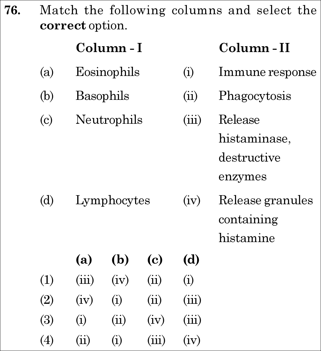
NEET 2020 · Q76 Easy
Official NEET 2020 Question 76 - Body Fluids and Circulation
1 (iii) (iv) (ii) (i) ✓ Key
2 (iv) (i) (ii) (iii)
3 (i) (ii) (iv) (iii)
4 (ii) (i) (iii) (iv)
💡 Verified Solution & 30-Second Hack
✓
Verified Against NTA Final Answer Key
Option (1) : (a)-(iii), (b)-(iv), (c)-(ii), (d)-(i)
📚 Body Fluids and Circulation Easy
📋 Given Parameters & Problem Setup
Column I (Leukocytes) (a) Eosinophils, (b) Basophils, (c) Neutrophils, (d) Lymphocytes
Column II (Functions) (i) Immune response, (ii) Phagocytosis, (iii) Release histaminase, destructive enzymes, (iv) Release granules containing histamine
⚡

The 30-Second Exam Hall Shortcut: ⚡ "Neutro = Phagocyte; Baso = Histamine; Eosino = Histaminase; Lympho = Immune!"

⏱️ 25 seconds

Neutrophils are phagocytic cells (c->ii). Basophils secrete histamine, serotonin, heparin (b->iv). Eosinophils resist infections and release histaminase/destructive enzymes (a->iii). Lymphocytes (B and T) mediate immune responses (d->i). Combination is (iii), (iv), (ii), (i) -> Option 1 in 10 seconds!

1 Neutrophils and monocytes are phagocytic cells destroying foreign organisms -> (c)-(ii).
2 Basophils secrete histamine, serotonin, heparin in inflammatory reactions -> (b)-(iv).
3 Eosinophils resist infections, associated with allergic reactions, and release histaminase -> (a)-(iii).
4 Lymphocytes (B and T forms) are responsible for adaptive immune responses -> (d)-(i).
5 Mapping: (a)-(iii), (b)-(iv), (c)-(ii), (d)-(i) -> Option 1.
🛡️
Why You Can 100% Trust This Shortcut: NCERT Biology · Human Physiology: Body Fluids and Circulation (Formed Elements: Leukocytes)
📐

Step-by-Step Rigorous Workflow & Mathematical Proof

Step 1

Match Eosinophils

Eosinophils (2-3% of total WBCs) resist infections and are associated with allergic reactions; they contain granules releasing histaminase and destructive enzymes to combat parasitic infections. Thus, (a) matches (iii).

Step 2

Match Basophils

Basophils (0.5-1% of total WBCs) secrete histamine, serotonin, and heparin, and are involved in inflammatory reactions. Thus, (b) matches (iv).

Step 3

Match Neutrophils

Neutrophils (60-65% of total WBCs) are the most abundant phagocytic cells that engulf and destroy foreign microbes entering the body. Thus, (c) matches (ii).

Step 4

Match Lymphocytes

Lymphocytes (20-25% of total WBCs) occur in two major types—B and T cells—both of which are responsible for the immune responses of the body (humoral and cell-mediated immunity). Thus, (d) matches (i).

⭐

360° Option Elimination Engine Key Feature

Diagnose every choice: Know exactly why the right option works and why distractors fail.

4-Option Deep Scan
1 A
✓ 100% Correct
Verdict & Scientific Reason:

Accurate pairing across all four white blood cell varieties and their distinct functions.

💡
Why this is airtight: None; core hematology curriculum.
2 B
✗ Incorrect
Verdict & Scientific Reason:

Incorrect; confuses eosinophils with histamine releasers and basophils with immune response.

⚠️
Student Fallacy (Why candidates pick this trap): Swapping basophil and eosinophil functions.
3 C
✗ Incorrect
Verdict & Scientific Reason:

Incorrect; assigns immune response to eosinophils and phagocytosis to basophils.

⚠️
Student Fallacy (Why candidates pick this trap): Mispairing primary leukocyte roles.
4 D
✗ Incorrect
Verdict & Scientific Reason:

Incorrect; inverts neutrophil phagocytosis with eosinophil enzymatic release.

⚠️
Student Fallacy (Why candidates pick this trap): Confusing granulocyte subsets.
⚠️

NTA Trap Alert: Basophil (Histamine) vs Eosinophil (Histaminase)

Basophils SECRETE Histamine (promoting inflammation). Eosinophils SECRETE Histaminase (destroying/breaking down histamine to moderate allergic reactions).

🛡️ How to avoid the -1 mark: Look at the suffix: Basophil releases Histam-INE; Eosinophil releases Histamin-ASE (the enzyme)!
📖

Core NEET Syllabus Standard

Zoology · Body Fluids and Circulation Standard Benchmark
Core Scientific Principle Tested:

""

Syllabus Focus: Leukocyte Types and Functions: Eosinophils, Basophils, Neutrophils & Lymphocytes
✓ Core NEET UG Syllabus Anchor Syllabus & Academic Standards ↗

Solve 4 NEET Questions a Day — One per Subject

Cover all 1,140 official NEET questions in 285 days at your own pace. Progress is saved on your device. No login needed.